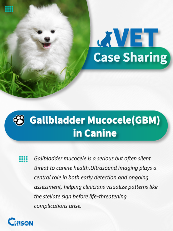
Breed: Pomeranian
Age: 4 years old
Gender: Male(neutered)
The gallbladder appeared moderately distended.
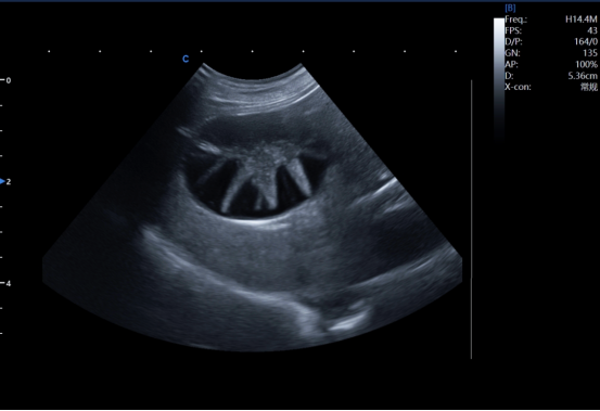
A radiating echogenic structure was observed within the lumen, forming a “stellate pattern”.
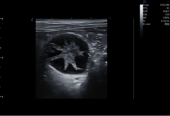
No signs of gallbladder wall thickening, perforation, or pericholecystic fluid were noted.
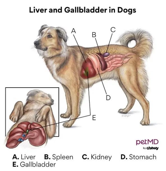
Thegallbladder is a small organ that stores and concentrates bile produced by theliver. Bile helps emulsify fats during digestion.
A GBM isan abnormal accumulation of thick, gelatinousmucus within the gallbladder, leading to its distension and oftenobstructing bile outflow.
While theexact pathogenesis is unclear, multiple factors are associated:
l Endocrine diseases, such as Cushing’s syndrome or hypothyroidism
l Hyperlipidemia
l Gallbladder dysmotility
l Genetic predisposition
GBM is more common in purebred dogs, especially:
l Miniature Schnauzers
l Shetland Sheepdogs
l Border Terriers
l Beagles
Left untreated, a GBM can lead to:
l Aseptic or septic cholecystitis
l Gallbladder wall necrosis
l Gallbladder rupture, causing bile peritonitis
l Systemic Inflammatory Response Syndrome (SIRS)
Early-stage GBMs may be asymptomatic, making imaging-based screening vital in at-risk dogs.
Four clinical outcomes of mucocele formation.
A—Gallbladder wall infarction and necrosis.
B—Secondary bacterial infection leading to superimposed cholecystitis.
C—Extrahepatic bile duct obstruction by mucus plugs.
D—Progressive accumulation of mucus leading to rupture under pressure.
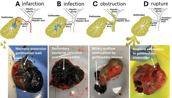
Picture from JAVMA I JUNE 2025| VOL 263| NO.6
Ultrasound is the gold standard for non-invasive diagnosis of GBM. It enables:
l Real-time visualization of gallbladder shape and internal content
l Identification of characteristic patterns and structural changes
l Early detection before rupture or infection develops
| Type | Description |
| Type I | Echogenic sludge occupying >30% of the lumen |
| Type II | Structured sludge with partial stellate strands |
| Type III | Distinct stellate pattern |
| Type IV | Mixed stellate and kiwi patterns |
| Type V | Kiwi pattern with echogenic debris |
| Type VI | Fully developed kiwi pattern, layered concentric structure |
l Early Stage: Mild mucin attachment to the gallbladder wall
l Immature Stage: Mucosal ridges projecting inward
l Mature Stage: Nearly complete obstruction of the lumen by mucin
l Overmature Stage: Dense, radiating echogenic streaks throughout the gallbladder
These changes are easily tracked over time using high-quality ultrasound, guiding decisions between conservative management and surgical intervention.
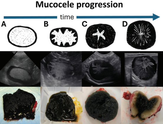
Simplified schematic of ultrasonographic progression of mucocele formation. A—Early mucocele formation: hypoechoic (black) rim of mucus attached to the gallbladder wall with central echogenic mobile or immobile sludge. B—Immature mucocele: ridges of hypoechoic mucus extending from and attached to the gallbladder wall with central echogenic mobile or immobile sludge. C—Mature mucocele:hypoechoic mucus attached to the gallbladder wall and nearly obliterating the gallbladder lumen with a small volume of central echogenic mobile or immobile sludge. D—Hypermature mucocele: highly compressed hypoechoic mucus obliterating the gallbladder lumen with fine hyperechoic striations radiating centrally.(Picture from JAVMA I JUNE 2025| VOL 263| NO.6)
Gallbladder mucocele is a serious but often silent threat to canine health.
Ultrasound imaging plays a central role in both early detection and ongoing assessment, helping clinicians visualize patterns like the stellate sign before life-threateningcomplications arise.
With advanced imaging technologies, CHISON VET is committed to empoweringveterinary professionals with clarity, precision, and care—supporting animal health, one scan at a time.
CHISONMEDICAL
References
1. Aguire AL, Center SA, Randolph JF, et al. Gallbladder disease in Shetland Sheepdogs: 38 cases (1995–2005). J Am Vet Med Assoc 231:79–88, 2007.
2. Amellem PM, Seim HB, MacPhail CM, et al. Long-term survival and risk factors associated with biliary surgery in dogs: 34 cases (1994–2004). J Am Vet Med Assoc 229:1451–1457, 2006.
3.Mehler SJ, Mayhew PD, Drobatz KJ and Holt DE. Variables associated with outcome in dogs undergoing extrahepatic biliary surgery: 60 Cases (1988–2002). Veterinary Surgery 33:644–649, 2004.
4.Pike FS, Berg J, King NW, et al. Gallbladder mucocele in dogs: 30 cases (2000–2002). J Am Vet Med Assoc 224:1615–1622, 2004.
5.Worley, DR, Hottinger Lawrence HJ. Surgical management of gallbladder mucoceles in dogs: 22 cases (1999–2003). J Am Vet Med Assoc 225:1418–1422, 2004.
6.Neer TM. A Review of disorders of the gallbladder and extrahepatic biliary Tract in the dog and cat. Journal of Veterinary Internal Medicine 6:186-192,1992.
7.Gall Bladder Mucocele in the Dog- Diagnosis and Medical Management,by Julie Stegeman, DVM, DACVIM, Nashville Veterinary Specialists - Clarksville
8.Canine Gallbladder Mucocele,BySharon A. Center, DVM, DACVIM, Department of Clinical Sciences, College of Veterinary Medicine, Cornell University
9.Diagnosis and management of gallbladder mucocele formation in dogs Jody L. Gookin
10.Diagnosis and management of gallbladder mucocele
formation in dogs, Jody L. Gookin, DVM, PhD, DACVIM1* ; Kyle G. Mathews, DVM, MS, DACVS; Gabriela Seiler, DVM, DACVR

Contact Us
Copyright © CHISON Medical Technologies Co., Ltd. All Rights Reserved | Privacy and terms of use
Products: Vascular Ultrasound Machine MSK Ultrasound Machine Portable Ultrasound Device Portable Ultrasound Machine for Sale Portable Ultrasound Machine For Pregnancy Handheld Ultrasound For Pregnancy Handheld Veterinary Ultrasound Veterinary Ultrasound Machine Portable Vascular Ultrasound Livestock Ultrasound Machine

CHISON respects your privacy. We use cookies to make our site more personal and enhance your experience. Read our Private Policy to learn more about cookies and how to manage them. You agree to our use of these technologies when you visit our site.

We appreciate your feedback
We sincerely invite you to participate in our survey for helping us to improve our digital market.
*1.How fast does the website load?
*2.Does the products displayed on the website interest you?
*3.How easy is it for you to find the information you need?
4.What information or service do you suggest we can offer?


THANKS
Thank you for sharing your thoughts with us. We’re highly appreciated your every feedback.

Sorry!

THANKS
Thank you for sharing your thoughts with us. We’re highly appreciated your every feedback.