Abstract:
Myxomatous Mitral Valve Disease (MMVD) in Canine is a common heart disease in elderly small dogs, characterized by fibrous thickening and junctional adhesion of the mitral valve closure margin. It is the most common acquired heart disease in canine, accounting for about 75% of clinical heart cases. Its main cause is progressive mucinous tumor-like lesions in the mitral valve. It will cause valve structure get thickening, prolapse, curling, and even chordae tendineae rupture, resulting in mitral insufficiency, mitral regurgitation, and ultimately leading to congestive heart failure in the left heart.
Pekingese, 10 years old, Female
Details: The dog suffered from wheezing and coughing, mainly dry cough, especially severe in the morning and evening. At the same time, it had poor appetite, with fatigue and disinterest in movement.
When performing ultrasound scanning, first take the right parasternal long axis section and the left apical four chamber view for examination, to determine the changes in the shape and structure of the mitral valve.
Then, the right parasternal short axis aortic section was taken to measure the ratio of the left atrium to the aorta, and the papillary muscle section was taken for M-mode echocardiography to measure the fractional shortening (FS).
Finally, continuous wave doppler was used to measure the regurgitation velocity at the mitral valve in the left apical four chamber view.
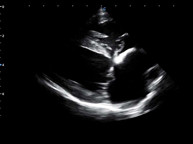
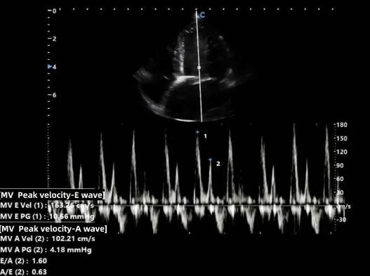
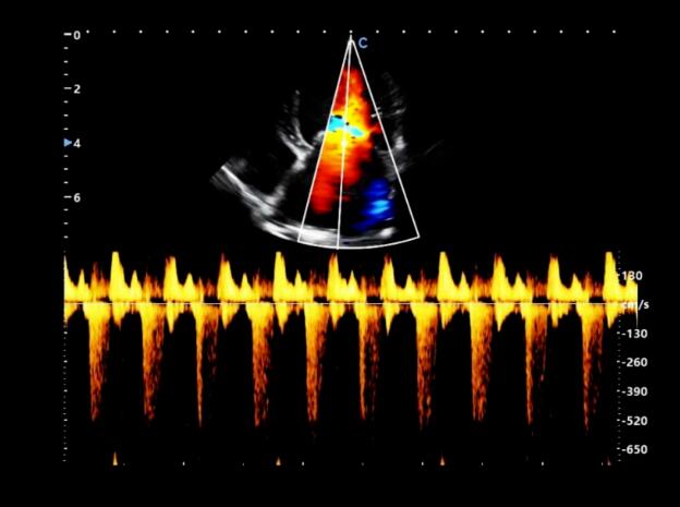
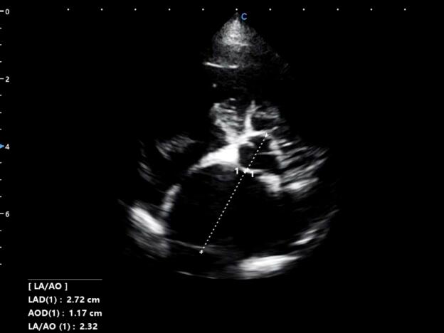
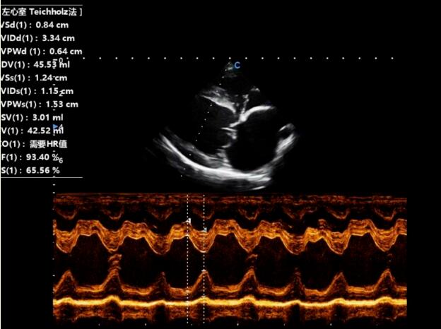
The examination results of the dog's heart showed that, MMVD caused mitral regurgitation, and left ventricular systolic retrograde mottled blood flow.
Based on the basic condition of this dog and the examination results of clinical, hematological, and imaging, it is diagnosed that the dog is afflicted with MMVD.
MMVD usually takes a long time from mild heart murmurs detected on initial examination to the heart failure at the end of the disease.
In the cases of early phase, only systolic regurgitant heart murmurs may be heard without obvious clinical symptoms. As the disease progresses, the dog may present symptoms such as cough, asthma, and exercise intolerance, especially after exercise and excitement. The main cause is left atrial dilation and compression of the left bronchus caused by mitral regurgitation, followed by reactive cough caused by increased pulmonary artery pressure and pulmonary edema.
It should be noted that, the above clinical symptoms are not unique to heart disease. Small elderly dogs are often accompanied by tracheal collapse and chronic bronchitis, which can also cause coughing and breathing difficulties. Clinical doctors need to make a differential diagnosis through medical history and other examinations.
Considering factors such as the age, vital signs, and breed of the sick dog, the treatment principle is chosen to be alleviating the cause and reduce the cardiac load. At the same time, the owner of the sick dog is advised to strengthen daily care and management and improve its quality of life, to extend its lifespan.
Echocardiography is the most sensitive method for diagnosing MMVD. Through it, we can visually observe the imaging changes of the mitral valve structure and evaluate the intensity of reflux and the function of the heart. On echocardiography, MMVD are commonly observed with varying degrees of dilation of the left atrium and left ventricle, and the thickness of the heart wall mostly normal or slightly increased. During the contraction period, thickening of one or two leaflets , and tumor like lesions at the end of the leaflets can be observed, and in severe cases, the leaflets may also be shortened.
Doppler echocardiography is very intuitive for diagnosing mitral regurgitation. The size and depth of its reflux also indicate the severity of the reflux.
Contact Us
Copyright © CHISON Medical Technologies Co., Ltd. All Rights Reserved | Privacy and terms of use
Products: Vascular Ultrasound Machine MSK Ultrasound Machine Portable Ultrasound Device Portable Ultrasound Machine for Sale Portable Ultrasound Machine For Pregnancy Handheld Ultrasound For Pregnancy Handheld Veterinary Ultrasound Veterinary Ultrasound Machine Portable Vascular Ultrasound Livestock Ultrasound Machine

CHISON respects your privacy. We use cookies to make our site more personal and enhance your experience. Read our Private Policy to learn more about cookies and how to manage them. You agree to our use of these technologies when you visit our site.

We appreciate your feedback
We sincerely invite you to participate in our survey for helping us to improve our digital market.
*1.How fast does the website load?
*2.Does the products displayed on the website interest you?
*3.How easy is it for you to find the information you need?
4.What information or service do you suggest we can offer?


THANKS
Thank you for sharing your thoughts with us. We’re highly appreciated your every feedback.

Sorry!

THANKS
Thank you for sharing your thoughts with us. We’re highly appreciated your every feedback.