Granulomatous mastitis (GM) is a chronic inflammatory disease occurring in the lobules of the mammary gland, first reported in 1972 by Kessler et al. It is clinically rare, occurring in only 1.8% of benign breast diseases.
The etiology of GM is unclear, and it is generally considered to be a rare chronic aseptic inflammatory breast disease, the incidence of which has been increasing year by year in China in recent years.
1. Elevated serum lactotide (antipsychotic about etc.).
2. Infectious factors.
3. Autoimmune factors.
4. Others: oral contraceptive pills, trauma, and so on.
1. Patients with granulomatous mastitis often consult the doctor for the sudden discovery of a lump, unilateral breast involvement is common, or both breasts at the same time or successive occurrence.
2. The lump is hard to palpation, but may become soft when an abscess forms, and may be accompanied by inflammatory enlargement of the axillary lymph nodes on the same side.
3. It mostly occurs in the peripheral part of the breast and develops to the center, which may involve the whole breast.
4. Generally accompanied by pain and skin redness and swelling, in severe cases, there may be ulceration and sinus tract formation, but also nipple overflow, nipple deformation, or inversion.
5. About 19% of cases of granulomatous lobular mastitis will present with nodular erythema or arthralgia of the upper and lower extremities.
The sonographic features of granulomatous mastitis vary according to the extent and degree of involvement of the lesion. Based on the image characteristics, the ultrasonographic manifestations of granulomatous mastitis have been classified and described in different ways.
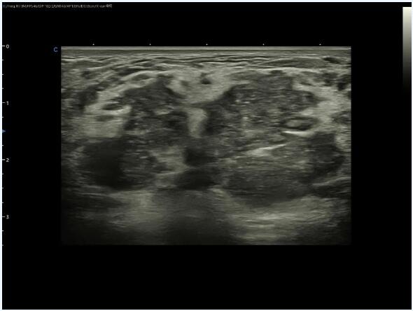
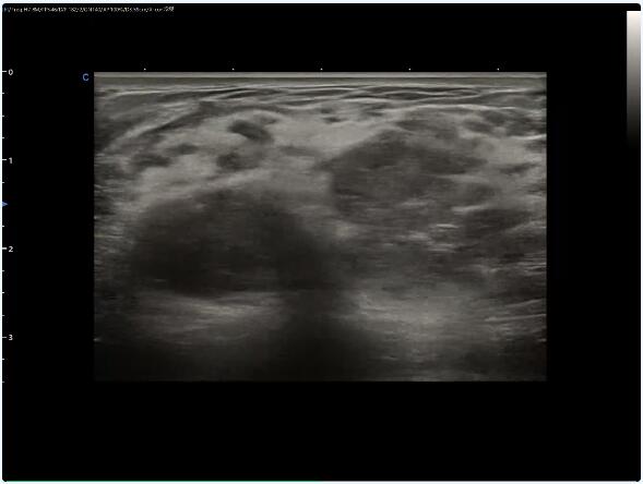
1. Mixed echogenic nodular type is mainly characterized by irregular shape of the mass, unclear boundary, hyperechoic ring around the mass, internal echogenicity is not homogeneous, and multiple scattered distribution of anechoic areas; lamellar hypoechoic type is mainly characterized by scattered distribution of anechoic areas within the lamellar hypoechoic area, and hyperechoic ring around the lesion, with some areas of unclear boundary; diffuse type is mainly characterized by diffuse distribution of hypoechoic and anechoic areas within the echogenicity of swollen glands; and diffuse distribution of hypoechoic and anechoic echoes within the echogenicity of swollen glands. The diffuse type is mainly characterized by a diffuse distribution of hypoechoic and anechoic areas in the echoes of the swollen gland.
2. The tubular type is characterized by a hypoechoic, heterogeneous mass surrounded by more hypoechoic ductal continuity; the solid type is characterized by a hypoechoic, heterogeneous mass surrounded by lobulation and angularity; and the diffuse type is characterized by a large patchy, diffuse glandular echogenicity that involves more than two quadrants, with no obvious localizing effect. Color Doppler ultrasound for benign and malignant lymph nodes helps in diagnosis and differential diagnosis. In patients with granulomatous mastitis, the axillary lymph nodes tend to be around 1.0 cm in long diameter, with an aspect ratio of <2. Blood flow is seen at the lymphatic gates in the form of thin strips or stellate dots, which is different from malignant enlarged lymph nodes.


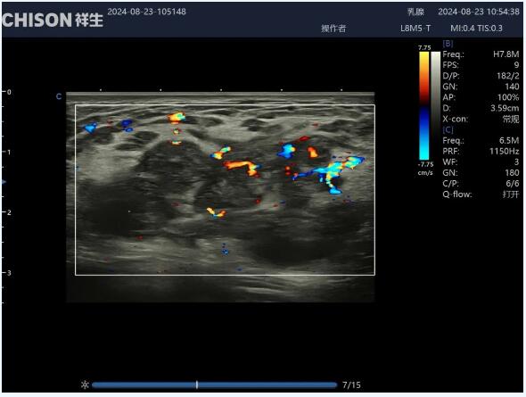
Patchy hypoechoic area is seen in the gland, and anechoic area is seen in the inner part of the gland, which is scattered, and a hyperechoic halo is seen around the lesion, which is thicker, with unclear boundary in some areas, and the internal blood flow signal is more abundant.
The ultrasound images of some granulomatous mastitis (mainly nodular) are very similar to those of invasive breast cancer, which is easy to misdiagnose, and the misdiagnosis rate has been reported to be 47%-63%. The main points of differentiation between the two are as follows:
1. Some granulomatous mastitis can be seen in the scattered distribution of echogenic areas, while invasive breast cancer echogenicity is rare, and the range is small and limited.
2. The hyperechoic halo of granulomatous mastitis is thicker and more diffuse, showing edema-like changes; while the hyperechoic halo of invasive breast cancer is thinner and more often combined with the burr sign, which is caused by the invasion of the mass into the peripheral tissues.
3. Although the morphology of granulomatous mastitis is irregular, it grows parallel to the gland, and the posterior echogenicity remains unchanged, while invasive breast cancer grows vertically, and the posterior echogenicity is often attenuated.
4. Color Doppler is also helpful in the differential diagnosis of granulomatous mastitis and invasive breast cancer. In the former, the internal blood flow is natural in shape, uniform in thickness, and low in resistance, while in the latter, the internal blood flow is irregular in shape, variable in thickness, and high in velocity and high in resistance.
5. Clinical manifestations are also helpful in the differential diagnosis of the two.
At present, there is no standardized treatment for granulomatous mastitis. There are mainly two kinds of surgical treatment and non-surgical treatment:
1. The former mainly involves incision and drainage of the abscess, lumpectomy, or extended excision;
2. The latter includes hormone therapy, methotrexate, and traditional Chinese medicine.
Contact Us
Copyright © CHISON Medical Technologies Co., Ltd. All Rights Reserved | Privacy and terms of use
Products: Vascular Ultrasound Machine MSK Ultrasound Machine Portable Ultrasound Device Portable Ultrasound Machine for Sale Portable Ultrasound Machine For Pregnancy Handheld Ultrasound For Pregnancy Handheld Veterinary Ultrasound Veterinary Ultrasound Machine Portable Vascular Ultrasound Livestock Ultrasound Machine

CHISON respects your privacy. We use cookies to make our site more personal and enhance your experience. Read our Private Policy to learn more about cookies and how to manage them. You agree to our use of these technologies when you visit our site.

We appreciate your feedback
We sincerely invite you to participate in our survey for helping us to improve our digital market.
*1.How fast does the website load?
*2.Does the products displayed on the website interest you?
*3.How easy is it for you to find the information you need?
4.What information or service do you suggest we can offer?


THANKS
Thank you for sharing your thoughts with us. We’re highly appreciated your every feedback.

Sorry!

THANKS
Thank you for sharing your thoughts with us. We’re highly appreciated your every feedback.