



Clinical Insights
Grayscale and color Doppler findings in a 49-year-old patient with bilateral peritesticular fluid, more prominent on the right.
Patient | 49-year-old man |
Examination | Scrotal ultrasound |
System | CHISON SonoMax |
Imaging Modes | 2D grayscale ultrasound and color Doppler flow imaging |
Scrotal swelling, a feeling of heaviness, or localized discomfort may involve the testes, epididymides, tunica vaginalis, spermatic cords, scrotal wall, or inguinal canal. A complete scrotal ultrasound examination should answer three practical questions: Where is the fluid? Is the testicular parenchyma abnormal? Is blood flow affected?
In this testicular ultrasound hydrocele case, both testes showed clear, homogeneous parenchymal echotexture. Anechoic fluid was seen around the testes and epididymides on both sides and was more prominent on the right. Color Doppler ultrasound demonstrated intratesticular blood flow, providing additional information for characterizing the finding and evaluating associated abnormalities.
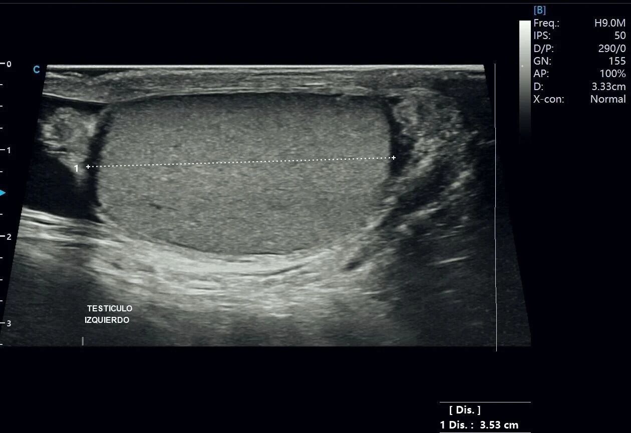
Figure 1. Longitudinal grayscale image of the left testis.
The left testis has a regular contour and measures approximately 3.53 cm in length in the displayed plane. The parenchymal echotexture is homogeneous and fine, with no definite focal abnormality visible in this image. A thin anechoic fluid layer surrounds the testis and is clearly separated from the testicular parenchyma.
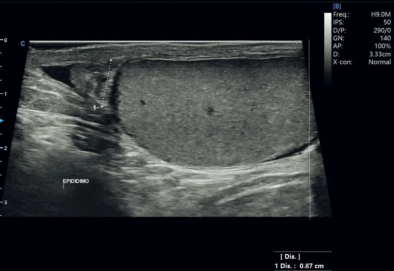
Figure 2. Grayscale image of the left epididymal head.
The left epididymal head is clearly visualized and measures approximately 0.87 cm along the displayed line. Its margins are relatively well defined, and the internal structure can be distinguished. Anechoic fluid is visible around the epididymis and adjacent testis, contrasting with the surrounding soft tissues.
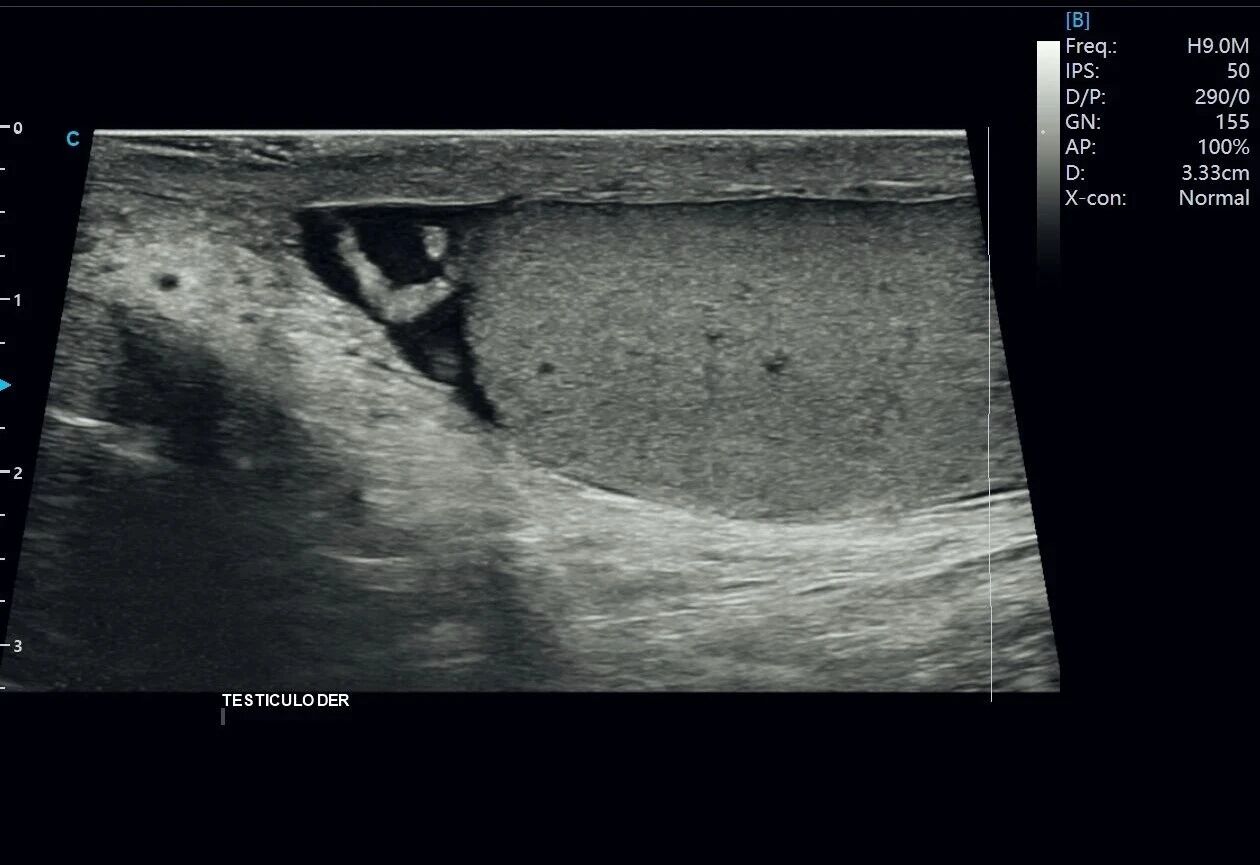 Figure 3. Grayscale image of the right testis with surrounding hydrocele.
The right testis has an intact contour and homogeneous parenchymal echotexture, with no definite focal solid lesion visible in the displayed plane. Anechoic fluid is present above and around the testis, outlining the interface between the testis and adjacent tissues. These are representative hydrocele ultrasound findings.
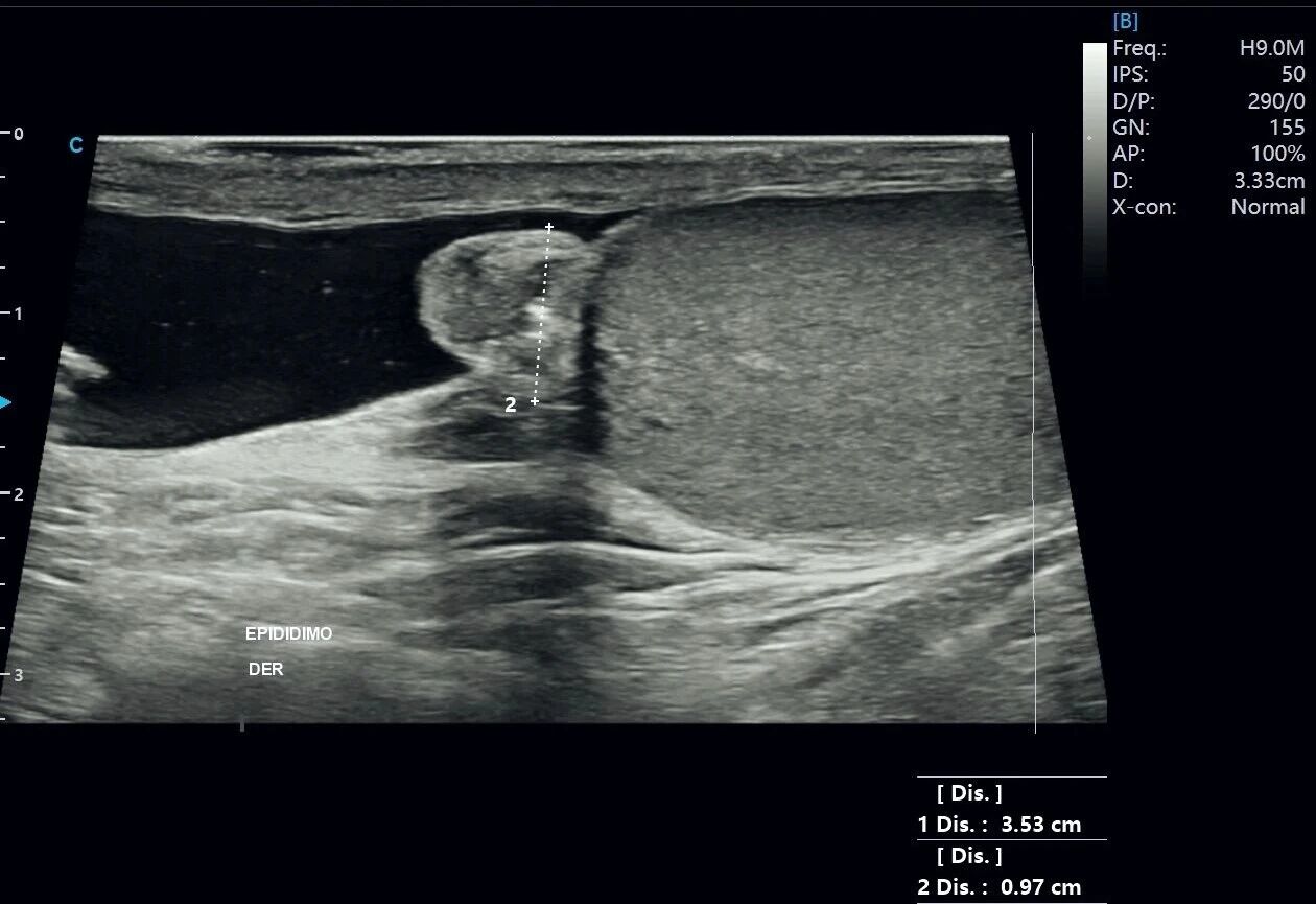 Figure 4. Grayscale image showing the right testis, epididymis, and surrounding fluid.
The right testis measures approximately 3.53 cm in length in the displayed plane, and the measured epididymal dimension is approximately 0.97 cm. The testicular echotexture is homogeneous, and the epididymal margins and internal structure are clearly shown. A relatively wide anechoic fluid collection with good acoustic transmission surrounds the testis and epididymis. Among the testicular ultrasound images in this case, this view most clearly demonstrates the spatial relationship between the testis, epididymis, and hydrocele.
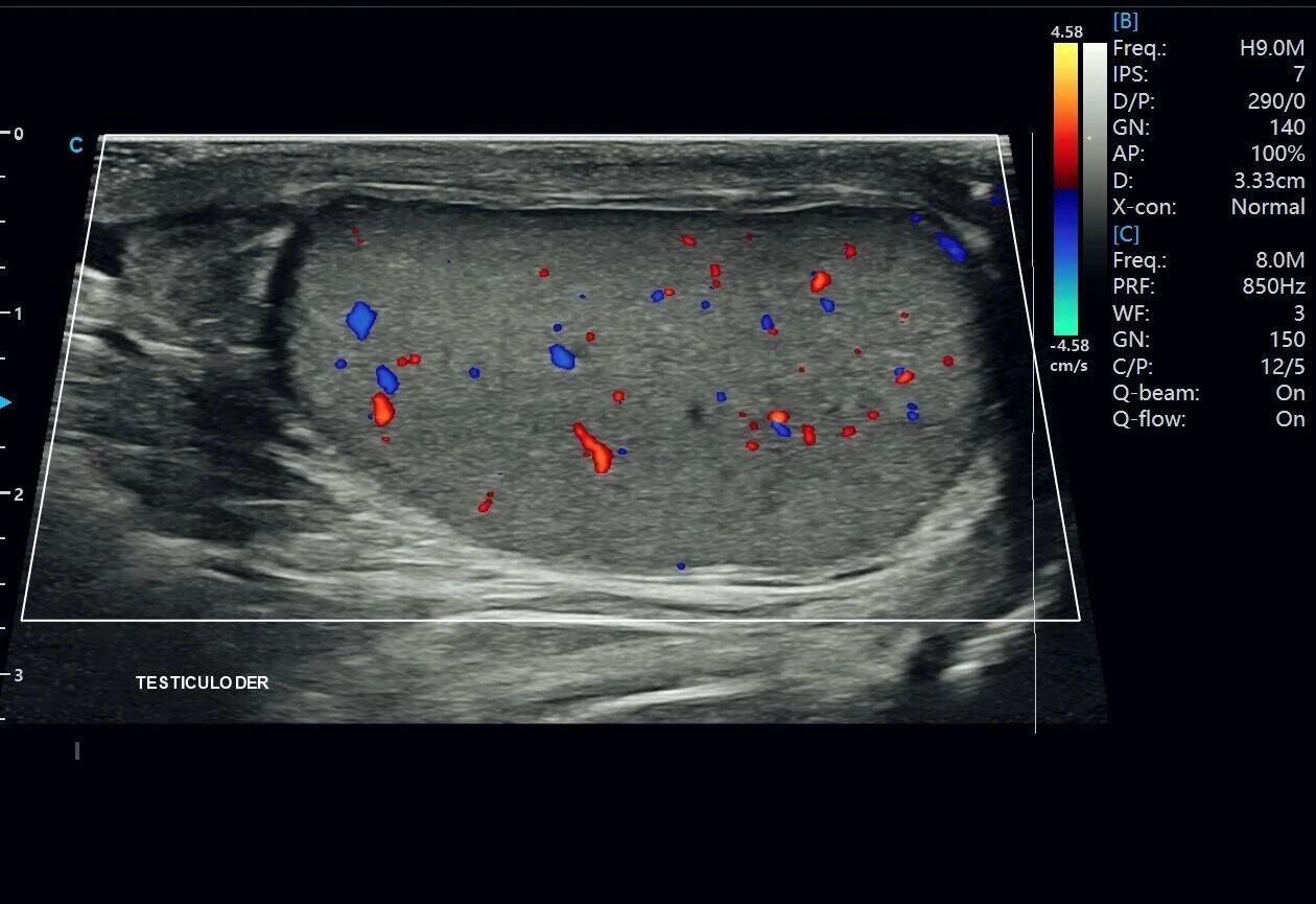
Figure 5. Color Doppler image of the right testis.
Color Doppler demonstrates multiple red and blue intratesticular flow signals, including small low-velocity signals. The testicular parenchyma remains homogeneous, the epididymal margin is clear, and anechoic fluid is visible around the testis. Color Doppler is used to assess blood flow in the testis and epididymis rather than flow within the fluid collection itself.
A hydrocele is most directly recognized on ultrasound as fluid within the tunica vaginalis surrounding part or all of the testis. A small amount of physiologic fluid may normally be present. When the volume increases and forms a visible fluid collection, hydrocele should be considered in the imaging assessment.
A simple hydrocele typically appears as an anechoic collection with good acoustic transmission. Fine internal echoes, septations, or dependent debris may suggest a chronic or complex collection, including possible hemorrhagic or infectious contents. Interpretation should be based on the full examination and clinical context.
In this case, the testicular parenchyma does not show a definite focal abnormality in the provided images. The key finding is the extratesticular fluid surrounding the testes, particularly on the right. This illustrates why scrotal ultrasound should not focus on the testes alone. The epididymides, tunica vaginalis, spermatic cords, scrotal wall, and other relevant extratesticular structures should also be assessed systematically, consistent with the AIUM practice parameter for scrotal ultrasound examinations.
Several other conditions may also appear as fluid-containing or cystic structures within the scrotum and should be considered when reviewing testicular ultrasound images:
· Epididymal cyst or spermatocele: usually appears as a localized cystic structure within or near the epididymal head, with a definable wall. It does not typically form a broad fluid collection around the testis as seen in this case.
· Hematocele or pyocele: may contain low-level echoes, fibrinous strands, septations, or debris rather than completely simple anechoic fluid. Scrotal wall thickening and inflammatory hyperemia may also be present, depending on the cause.
· Inguinoscrotal hernia: assessment should determine whether the finding is continuous with the inguinal canal. Bowel loops, fat, or peristalsis may be visible instead of a simple fluid collection surrounding the testis.
The combination of fluid morphology, anatomic compartment, and the appearance of the testes and epididymides is central to characterizing scrotal fluid collections. Grayscale ultrasound helps localize a finding as intratesticular or extratesticular, while color Doppler adds information about tissue perfusion.
· Hydrocele fluid is extratesticular and located within the tunica vaginalis; on ultrasound, it commonly appears as an anechoic collection partly or completely surrounding the testis.
· Simple fluid generally transmits sound well. Septations, internal echoes, strands, or debris may indicate a more complex collection that requires correlation with the clinical context.
· After identifying fluid, examine the testes and epididymides for contour, echotexture, focal lesions, and blood flow to assess for possible associated causes or complications.
· Color Doppler evaluates blood flow in the testes and epididymides, not the hydrocele itself. In this case, small intratesticular low-velocity flow signals were clearly displayed with SonoMax.
This case demonstrates how a structured grayscale and color Doppler examination can show the location and character of scrotal fluid while supporting assessment of the testes, epididymides, and intratesticular blood flow. SonoMax provided clear anatomical and Doppler information for this testicular hydrocele ultrasound evaluation.
1. Hoang VT, Van HAT, Hoang TH, et al. A Review of Classification, Diagnosis, and Management of Hydrocele. J Ultrasound Med. 2024;43(3):599-607. doi:10.1002/jum.16380.
2. Patil V, Shetty SMC, Das S. Common and Uncommon Presentation of Fluid within the Scrotal Spaces. Ultrasound Int Open. 2015;1(2):E34-E40. doi:10.1055/s-0035-1555919.
3. American Institute of Ultrasound in Medicine. AIUM Practice Parameter for the Performance of Scrotal Ultrasound Examinations, 2025 Revision. J Ultrasound Med. 2025. doi:10.1002/jum.70127.
4. Pilatz A, Rusz A, Wagenlehner F, et al. Reference values for testicular volume, epididymal head size and peak systolic velocity of the testicular artery in adult males measured by ultrasonography. Ultraschall Med. 2013;34(4):349-354.
Disclaimer: For professional education and information only. This article does not replace clinical judgment or medical care. If symptoms are present, seek assessment from a qualified healthcare professional. Some source materials and images were compiled from publicly available resources; please contact CHISON regarding any rights concerns.

Contact Us
Copyright © CHISON Medical Technologies Co., Ltd. All Rights Reserved | Privacy and terms of use
Products: Vascular Ultrasound Machine MSK Ultrasound Machine Portable Ultrasound Device Portable Ultrasound Machine for Sale Portable Ultrasound Machines For Pregnancy Handheld Ultrasound For Pregnancy Handheld Veterinary Ultrasound Veterinary Ultrasound Machine Portable Vascular Ultrasound Livestock Ultrasound Machine

CHISON respects your privacy. We use cookies to make our site more personal and enhance your experience. Read our Private Policy to learn more about cookies and how to manage them. You agree to our use of these technologies when you visit our site.
We appreciate your feedback
We sincerely invite you to participate in our survey for helping us to improve our digital market.
*1.How fast does the website load?
*2.Does the products displayed on the website interest you?
*3.How easy is it for you to find the information you need?
4.What information or service do you suggest we can offer?

THANKS
Thank you for sharing your thoughts with us. We’re highly appreciated your every feedback.
Sorry!
THANKS
Thank you for sharing your thoughts with us. We’re highly appreciated your every feedback.
