


Clinical Insights
Liver Ultrasound: What It Shows, Benefits and Elastography
A liver ultrasound uses sound waves to create real-time images of the liver and nearby structures. Also called liver sonography, liver echography, or a sonogram of the liver, it is commonly included in an abdominal ultrasound examination and does not use ionizing radiation.
B-mode ultrasound shows structure, Doppler evaluates blood flow, and shear wave elastography estimates tissue stiffness. These techniques answer different questions, and qualified professionals interpret the findings together with symptoms, laboratory results, medical history, and other tests when needed.
During a liver ultrasound procedure, a trained sonographer or clinician moves a transducer over the upper abdomen. The system converts returning sound-wave echoes into images viewed in real time. A routine scan begins with two-dimensional B-mode imaging and may also include the gallbladder, bile ducts, abdominal vessels, Doppler measurements, or elastography depending on the referral question.
A healthcare provider may order an abdominal ultrasound for the liver because of symptoms, abnormal laboratory results, a known condition, or a need for follow-up. Common reasons include:
· Symptoms or examination findings, such as abdominal pain, swelling, or a suspected enlarged liver.
· Laboratory abnormalities that require imaging correlation.
· Possible diffuse or focal changes, including fatty change, chronic liver disease, a cyst, mass, bile-duct dilatation, or surrounding fluid.
· Blood-flow assessment with Doppler ultrasound or follow-up of a previous finding.
The clinical question determines the protocol. Ultrasound supports evaluation but may need to be complemented by laboratory tests, CT, MRI, elastography, biopsy, or specialist review.
A liver ultrasound can show organ size, contour, echotexture, focal findings, bile ducts, surrounding fluid, and visible vessels. Routine imaging may show changes associated with fat or advanced chronic disease, but it cannot by itself determine inflammation, reliably stage all fibrosis, or provide a final diagnosis for every lesion. Doppler adds information about blood-flow direction and velocity in the portal and hepatic circulation.
B-mode creates two-dimensional grayscale images and forms the basis of most liver examinations. It supports assessment of size, contour, echogenicity, focal findings, bile ducts, and adjacent anatomy.
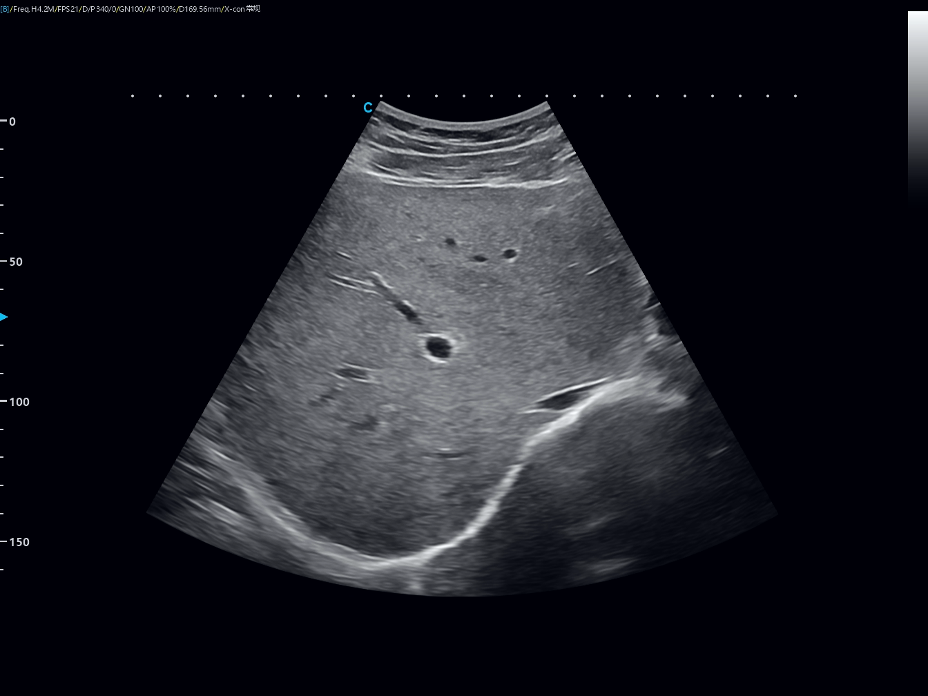
B-mode liver ultrasound image showing liver structure
Color Doppler displays the presence and direction of blood flow, while spectral Doppler provides waveforms and velocity measurements. It may be used for portal or hepatic circulation, suspected obstruction, transplant follow-up, or lesion vascularity.
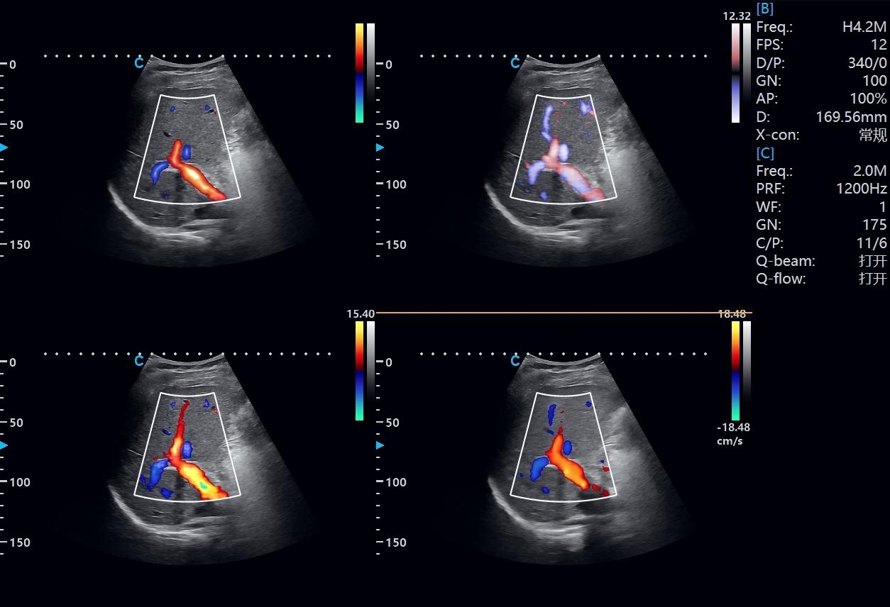
Color Doppler ultrasound assessment of hepatic blood flow
Ultrasound shear wave elastography estimates liver stiffness. Point shear wave elastography (P-SWE) measures a selected sampling region, while two-dimensional shear wave elastography (2D-SWE) displays a broader color-coded map. Increased stiffness can be associated with fibrosis, but inflammation, congestion, cholestasis, recent food intake, and acquisition quality may affect results. A liver ultrasound with elastography supplements B-mode imaging and clinical assessment.
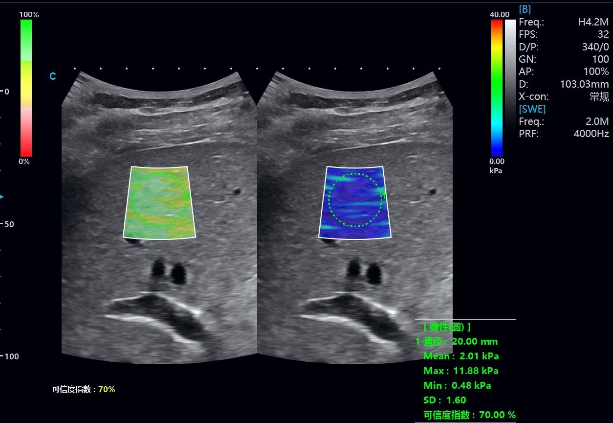
CHISON liver ultrasound with shear wave elastography
· No ionizing radiation. Diagnostic ultrasound uses sound waves rather than x-rays.
· Noninvasive, real-time imaging. The operator can adjust views and add Doppler or elastography when indicated.
· Flexible use. Examinations can be performed in imaging departments, clinics, and selected bedside settings.
· Follow-up and procedure support. Ultrasound can compare findings over time or guide selected interventions when performed by trained professionals.
Preparation varies by facility and protocol. Some patients are asked to avoid food or drink for several hours because bowel gas and gallbladder contraction can affect visualization; others need little preparation. Follow the instructions from the doctor or imaging center. During the scan, the operator may ask the patient to change position or briefly hold a breath.
A liver ultrasound is usually completed during a short appointment, but timing varies with the protocol, anatomy, patient condition, and whether Doppler, elastography, or a procedure is included. The facility can provide the most accurate estimate.
Bowel gas, depth, body habitus, and overlapping appearances can limit ultrasound. A normal scan does not exclude every form or stage of liver disease, and routine imaging cannot determine inflammation or prove steatohepatitis. Findings should be correlated with clinical and laboratory information, with further imaging or follow-up when recommended.
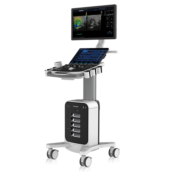
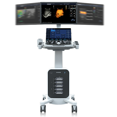
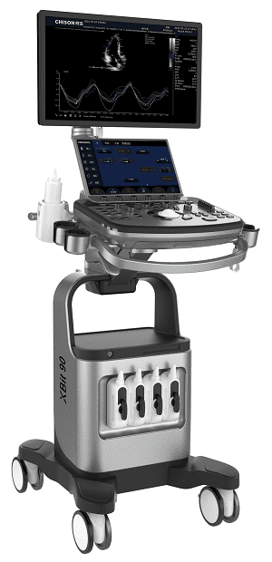
CHISON provides general-imaging systems for B-mode and Doppler examinations. Verified product pages for SonoMax 7, SonoMax 9, and XBit 90 also list P-SWE, 2D-SWE, velocity values, and Young’s modulus for selected configurations, supporting structural, blood-flow, and tissue-stiffness assessment.
CHISON system | Format | Relevant verified capabilities |
| Cart-based general imaging | P-SWE, 2D-SWE, velocity and Young’s modulus parameters |
| Cart-based general imaging | P-SWE, 2D-SWE, velocity and Young’s modulus parameters |
| Cart-based ultrasound | P-SWE, 2D-SWE, quantitative parameters, and Doppler workflow |
Functions, transducers, regulatory clearances, and intended uses vary by country and configuration. Explore CHISON general imaging and shear wave elastography solutions or contact a CHISON representative for local specifications.
Yes. Liver ultrasound, liver sonography, liver echography, and sonogram of the liver commonly refer to the same sound-wave-based imaging method. The protocol may include B-mode, Doppler, or elastography.
Routine ultrasound may show changes consistent with liver fat but cannot determine inflammation or reliably stage fibrosis. Shear wave elastography estimates stiffness as an additional marker, interpreted with laboratory and clinical findings.
Instructions vary by facility and examination. Follow the fasting, water, and medication directions provided by the ordering clinician or imaging center.
No. Some conditions may not produce visible changes on routine ultrasound. Results must be considered with symptoms, history, laboratory tests, and recommended follow-up.
Doppler evaluates blood flow, while elastography estimates tissue stiffness. They provide different information and may be included in the same examination.
Contact Us
Copyright © CHISON Medical Technologies Co., Ltd. All Rights Reserved | Privacy and terms of use
Products: Vascular Ultrasound Machine MSK Ultrasound Machine Portable Ultrasound Device Portable Ultrasound Machine for Sale Portable Ultrasound Machines For Pregnancy Handheld Ultrasound For Pregnancy Handheld Veterinary Ultrasound Veterinary Ultrasound Machine Portable Vascular Ultrasound Livestock Ultrasound Machine

CHISON respects your privacy. We use cookies to make our site more personal and enhance your experience. Read our Private Policy to learn more about cookies and how to manage them. You agree to our use of these technologies when you visit our site.
We appreciate your feedback
We sincerely invite you to participate in our survey for helping us to improve our digital market.
*1.How fast does the website load?
*2.Does the products displayed on the website interest you?
*3.How easy is it for you to find the information you need?
4.What information or service do you suggest we can offer?

THANKS
Thank you for sharing your thoughts with us. We’re highly appreciated your every feedback.
Sorry!
THANKS
Thank you for sharing your thoughts with us. We’re highly appreciated your every feedback.