


Global Expert Stories
How elastography and mobile vascular ultrasound support real-world care
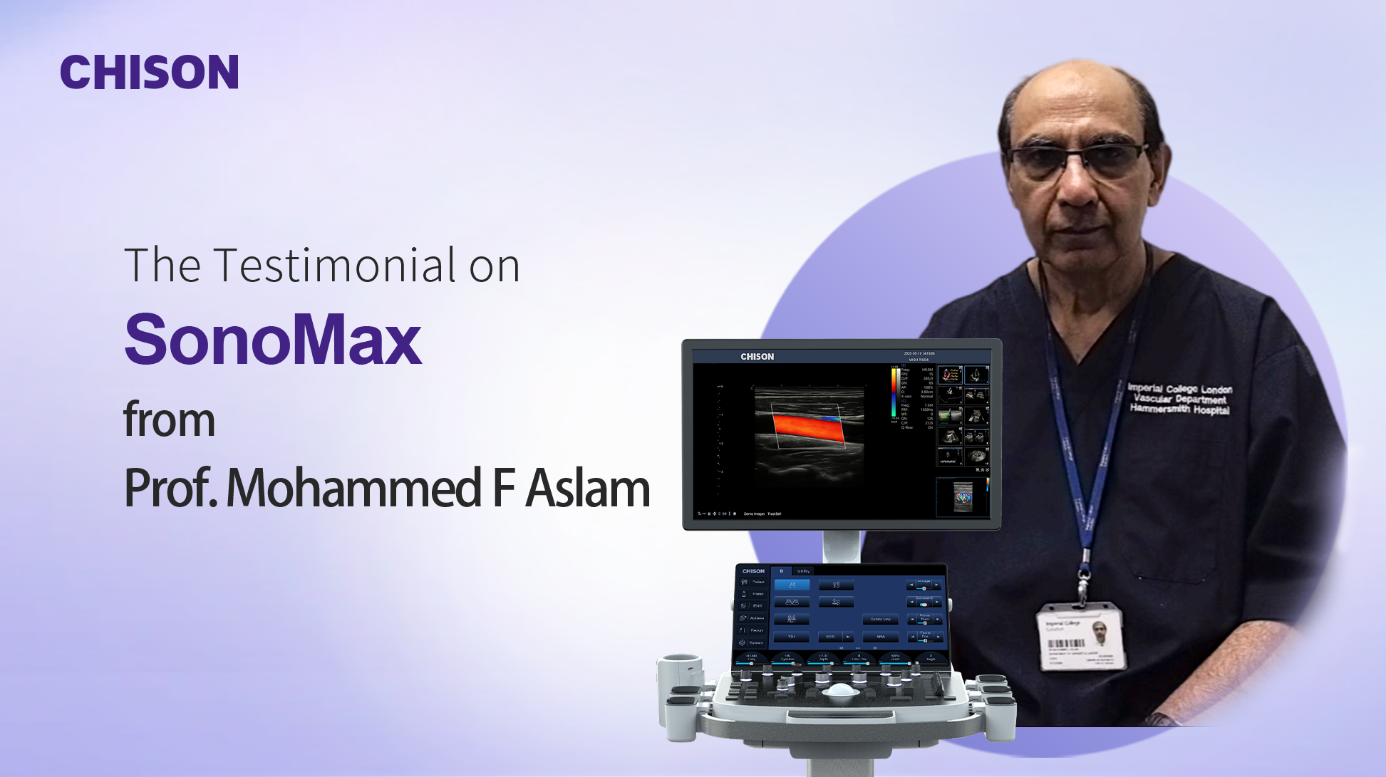
A clinical perspective based on Prof. Mohammed F. Aslam’s vascular ultrasound practice with SonoMax 9.
In vascular ultrasound, a clear image is only the beginning. The more important question is whether the examination provides enough information to guide the next clinical decision.
In a high-volume hospital environment, vascular ultrasound rarely follows a single, predictable path. One examination may take place in a renal unit, another in intensive care, and the next in an operating theatre. The clinical question also changes from patient to patient: Is a dialysis fistula failing? What is limiting blood flow? Does a thrombus appear acute or chronic? Is immediate intervention necessary, or would observation be safer?
These are the questions that shape the work of Prof. Mohammed F. Aslam, who leads a medical ultrasound programme and works within vascular surgery and circulatory sciences at Imperial College London and Imperial College Healthcare NHS Trust. With approximately two decades of experience in clinical practice and medical education, he has seen ultrasound technology evolve from an image-acquisition tool into a broader source of diagnostic and management information.
His experience with the CHISON SonoMax 9 is therefore best understood not as a product story, but as a clinical story: how mobility, vascular Doppler ultrasound, and elastography can help clinicians move from seeing an image to answering a patient-care question.
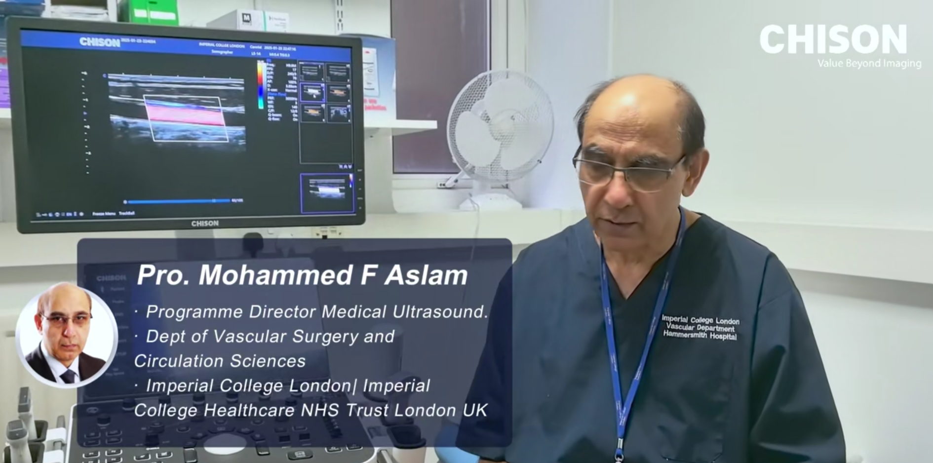
Prof. Mohammed F. Aslam discusses how advanced ultrasound technology can translate into practical clinical value.
The story of a vascular ultrasound examination does not begin with a feature list. It begins with uncertainty.
Consider a dysfunctional arteriovenous fistula in a patient receiving dialysis. A technically acceptable image may show the vessel, but the clinician still needs to understand what is happening within it. Where is the abnormality? How is blood moving through the access? Is there stenosis, altered flow, or another problem that could affect dialysis? The useful examination is the one that helps convert these findings into a management plan.
This is why Prof. Aslam places clinical relevance ahead of image quality alone. High-resolution vascular ultrasound matters, but its value depends on whether it provides reliable information about vascular structure, hemodynamics, and possible pathology. The image is evidence; the decision is the objective.
Before an examination can answer a clinical question, the system must reach the place where care is happening. In a hospital, that may mean moving between intensive care, an operating theatre, a renal service, and other departments during the same working day.
For Prof. Aslam, this is where a mobile ultrasound system becomes clinically meaningful. Rather than transporting a large conventional unit through multiple departments, the examination can be performed closer to the patient. This reduces logistical friction and allows vascular imaging to become part of the immediate workflow instead of a separate event that must be arranged elsewhere.
The transducer also matters. Lightweight, streamlined probes are easier to manipulate when space is limited or when the scan angle must be adjusted carefully. In vascular ultrasound, small changes in positioning can influence whether the clinician obtains the view or Doppler information needed to answer the question.
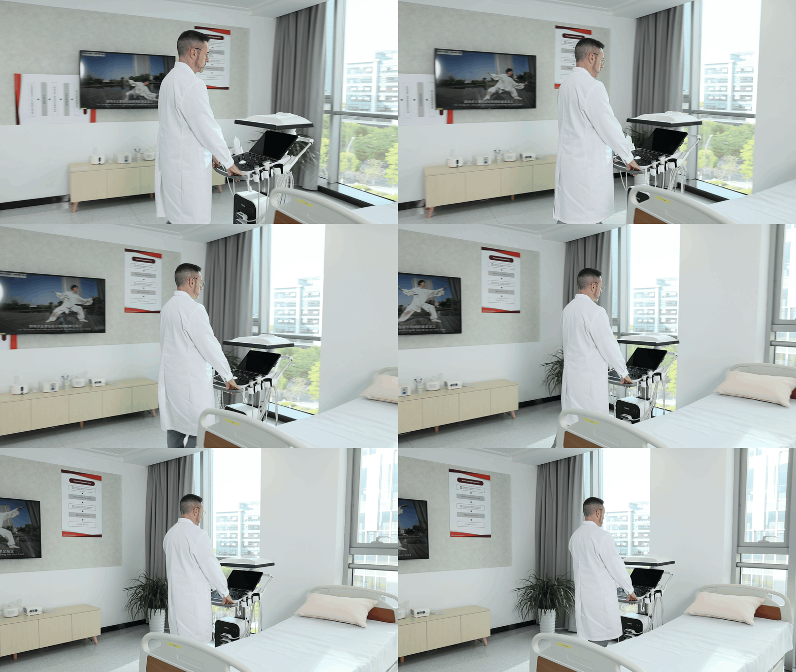
A mobile ultrasound system can support examinations in clinical environments where space, positioning, and access are constrained.
Vascular ultrasound is not only about the shape of a vessel. It is also about movement: the direction, pattern, and velocity of blood flow.
In Prof. Aslam’s workflow, optimized vascular imaging and Doppler analysis help build a more complete picture of the patient’s condition. SonoPW, for example, displays multiple spectral velocity measurements from different sites simultaneously in real time. This can help clinicians compare flow behavior across selected locations rather than interpreting each site in isolation.
For dialysis fistula ultrasound, this type of information can be especially relevant. A clinician may need to trace how blood moves through the access, identify areas of altered flow, and connect those findings with the patient’s clinical presentation. The purpose is not to generate more data for its own sake, but to make the hemodynamic story easier to understand.
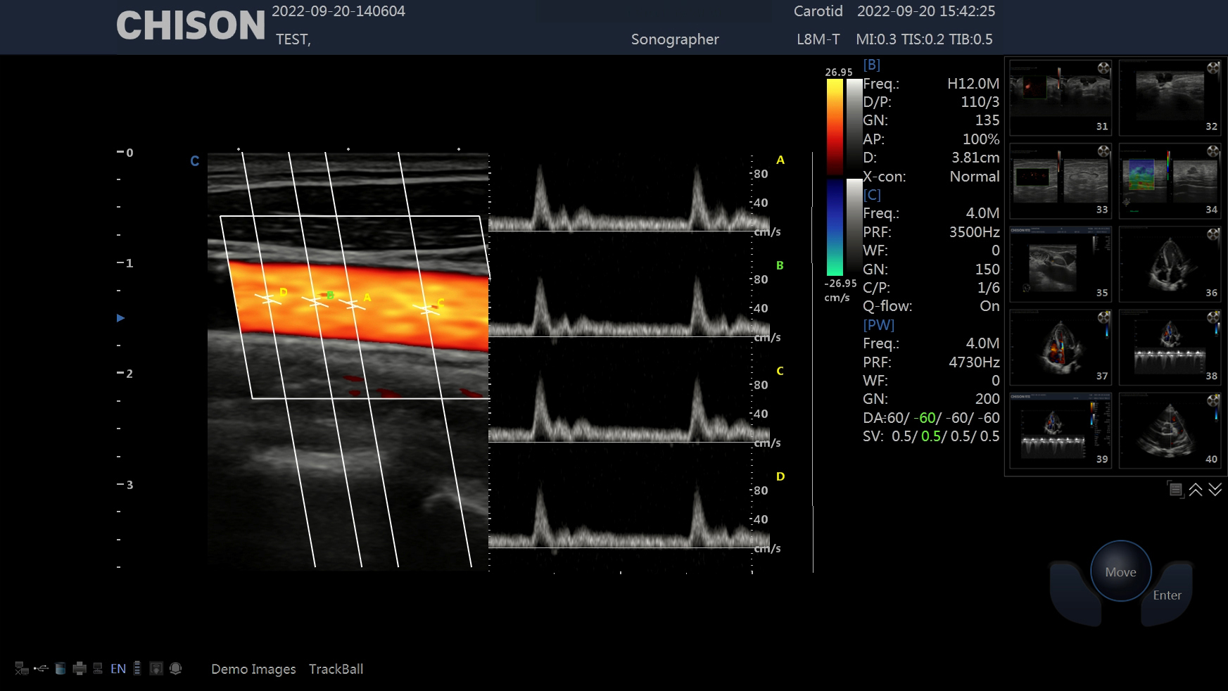
The above image depicts the SonoMax 9 ‘s unique SonoPW feature, which provides multiple spectral velocities at different sites simultaneously in real time
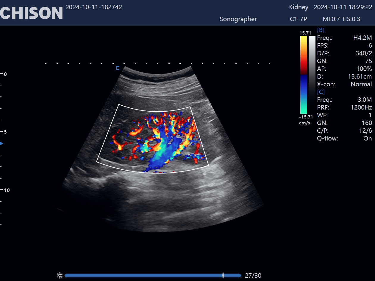
Renal Color Doppler : SonoMax 9
A thrombus creates another type of clinical uncertainty. The question is not simply whether thrombus is present. Clinicians may also need to consider whether it appears acute, chronic, or relatively recent, because that assessment can influence the urgency and type of treatment under consideration.
Conventional ultrasound findings remain central, but elastography and shear wave elastography may add information about tissue stiffness and thrombus characteristics. Used alongside vascular Doppler ultrasound, these techniques can contribute another layer of evidence when clinicians are trying to understand the condition of the thrombus.
That information may affect whether thrombolytic therapy is considered appropriate or whether observation and follow-up provide a more suitable path. The technology does not make the treatment decision. It can, however, help strengthen the information on which that decision is based.
The strongest theme in Prof. Aslam’s account is that advanced vascular ultrasound should be judged by what happens after the image appears on the screen.
Does the examination help the team identify why a fistula is dysfunctional? Does it provide enough information to characterize a thrombus more confidently? Can it help clinicians select an appropriate management pathway and potentially avoid an unnecessary intervention? Can the system move efficiently to the patient rather than forcing the patient and clinical team to adapt around the equipment?
These questions connect imaging performance with patient outcomes, workflow, and healthcare resource use. Treatments such as thrombolytic therapy involve clinical risk as well as cost. More complete ultrasound information may help clinicians make better-informed choices about when to intervene, when to monitor, and what additional assessment may be necessary.
In that context, SonoMax 9 is not presented as the hero of the story. It is one part of the clinical environment: a mobile platform through which vascular Doppler ultrasound, SonoPW, elastography, shear wave elastography, and image optimization can be applied to real clinical questions.
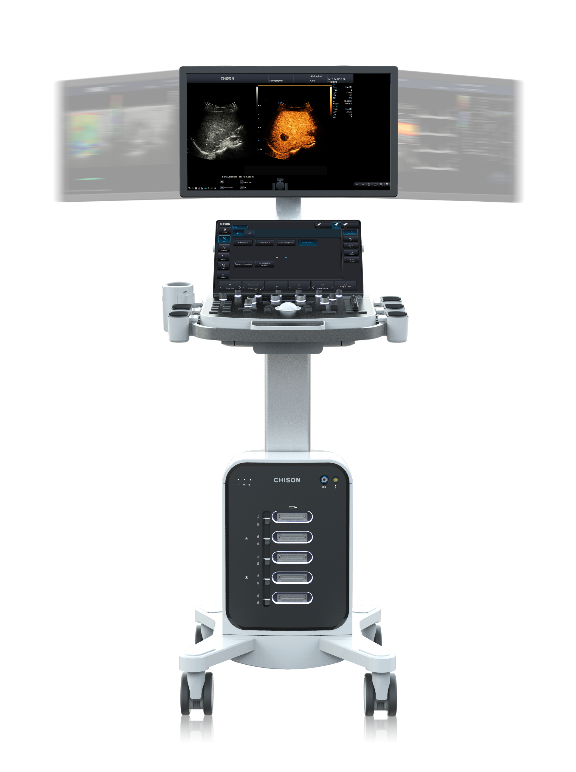
SonoMax 9 as part of a broader vascular ultrasound workflow focused on clinical decision support

Vascular ultrasound is used to evaluate blood vessels, vascular structure, and blood-flow patterns. In the clinical context described by Prof. Aslam, it supports applications such as dialysis fistula assessment, thrombus evaluation, and investigation of vascular abnormalities.
A vascular ultrasound can provide grayscale images of vessel anatomy together with Doppler information about the direction and velocity of blood flow. Depending on the examination and technology used, it may also provide additional information about tissue or thrombus characteristics.
Shear wave elastography provides information related to tissue stiffness. In vascular practice, it may complement conventional ultrasound and Doppler findings when clinicians are evaluating tissue or thrombus characteristics.
A mobile ultrasound system can be moved between intensive care, operating theatres, renal units, and other departments. This allows examinations to take place closer to the patient and reduces the logistical burden of moving larger equipment.
The evolution of vascular ultrasound is often described through new imaging modes and system specifications. Prof. Aslam’s experience offers a more useful way to understand that progress: follow the clinical question.
The examination may begin with a mobile system at the bedside, continue through Doppler analysis and elastography, and end with a decision about treatment, monitoring, or further investigation. What matters is not that the technology can do more, but that it helps the clinician understand more.
That is the shift from imaging to clinical decisions—and the point at which ultrasound technology begins to create meaningful value for patients.
Contact Us
Copyright © CHISON Medical Technologies Co., Ltd. All Rights Reserved | Privacy and terms of use
Products: Vascular Ultrasound Machine MSK Ultrasound Machine Portable Ultrasound Device Portable Ultrasound Machine for Sale Portable Ultrasound Machine For Pregnancy Handheld Ultrasound For Pregnancy Handheld Veterinary Ultrasound Veterinary Ultrasound Machine Portable Vascular Ultrasound Livestock Ultrasound Machine

CHISON respects your privacy. We use cookies to make our site more personal and enhance your experience. Read our Private Policy to learn more about cookies and how to manage them. You agree to our use of these technologies when you visit our site.
We appreciate your feedback
We sincerely invite you to participate in our survey for helping us to improve our digital market.
*1.How fast does the website load?
*2.Does the products displayed on the website interest you?
*3.How easy is it for you to find the information you need?
4.What information or service do you suggest we can offer?

THANKS
Thank you for sharing your thoughts with us. We’re highly appreciated your every feedback.
Sorry!
THANKS
Thank you for sharing your thoughts with us. We’re highly appreciated your every feedback.
