



HOT NEWS
Handheld ultrasound is changing where and how ultrasound imaging can be used. By bringing real-time imaging directly to the point of care, clinicians can use ultrasound for focused assessment, clinical decision support, procedure guidance and follow-up evaluation without relying exclusively on a dedicated ultrasound room.
This versatility is particularly valuable in emergency medicine, critical care, anesthesia, vascular access and other environments where fast access to anatomical information matters.
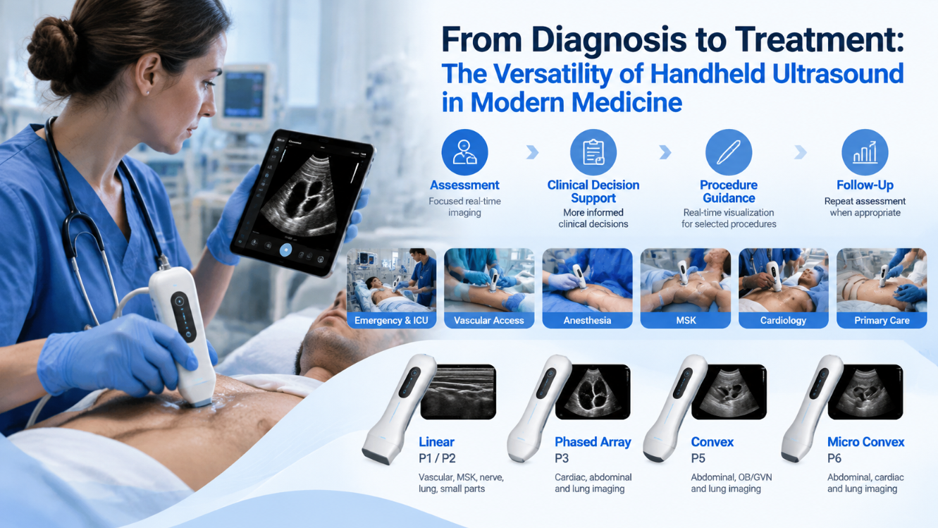
Point-of-care ultrasound (POCUS) is generally performed by healthcare professionals at or near the patient's bedside to answer focused clinical questions.
Depending on the probe and clinical application, handheld ultrasound can support assessment of areas such as:
Heart
Lungs
Abdomen
Blood vessels
Musculoskeletal structures
Nerves
Small and superficial structures
For example, a phased-array probe can support focused cardiac and lung examinations, while a linear probe is commonly suited to vascular, MSK, nerve and superficial imaging.
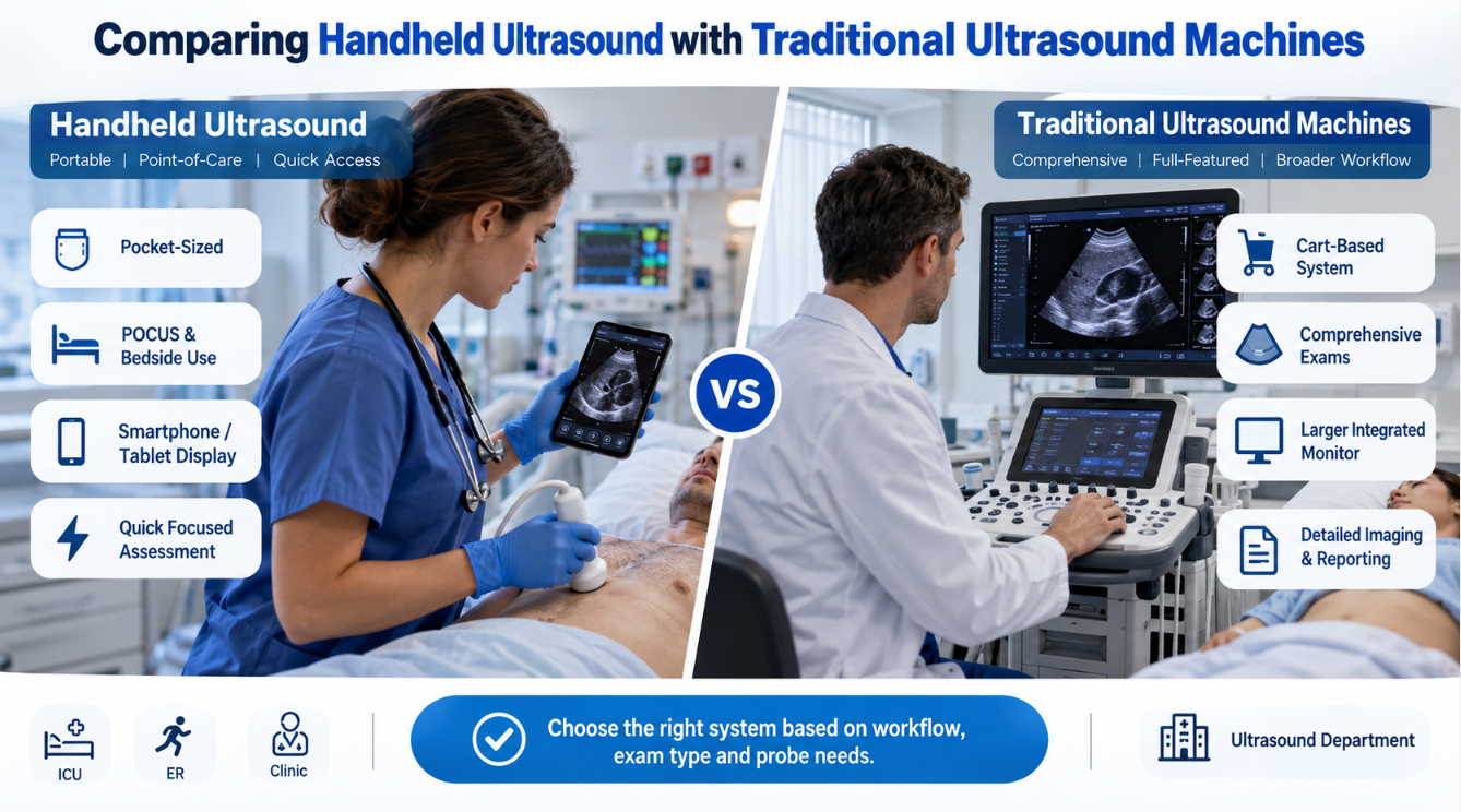
The purpose is not necessarily to replace a comprehensive ultrasound study. Instead, handheld ultrasound gives clinicians another imaging tool that can be integrated into the immediate clinical workflow.
The role of handheld ultrasound can extend beyond assessment.
Real-time visualization of anatomical structures can support ultrasound-guided procedures such as vascular access and selected needle-guided interventions. By seeing vessels, surrounding tissues and other relevant anatomy during a procedure, clinicians can use ultrasound as an additional source of visual guidance.
This creates a workflow in which the same portable imaging platform may support both initial assessment and subsequent procedural guidance.
Clinical Setting | Potential Role of Handheld Ultrasound |
Emergency Medicine | Focused cardiac, lung and abdominal assessment |
ICU & Critical Care | Bedside monitoring and focused examinations |
Vascular Access | Vessel visualization and needle guidance |
Anesthesia | Nerve and anatomical visualization for selected procedures |
MSK | Muscles, tendons, joints and superficial structures |
Cardiology | Focused cardiac ultrasound |
Primary Care | Focused examinations when immediate imaging is useful |
Mobile Care | Portable imaging where full-size equipment is difficult to deploy |
The exact application depends on the device, probe configuration, operator training and local clinical protocols.
No single probe is optimal for every examination. Probe selection should therefore follow the anatomy and clinical application.
CHISON offers several SonoEye handheld ultrasound configurations:
SonoEye P1 and P2 – Linear Probes
Designed for applications including vascular, MSK, nerve, lung and small-parts imaging. Their linear-array configuration makes them suitable for many superficial structures and ultrasound-guided applications.
SonoEye P3 – Phased Array
Designed for cardiac, abdominal and lung applications. The phased-array format is particularly relevant when scanning through smaller acoustic windows.
SonoEye P5 – Convex Probe
Designed for abdominal, OB/GYN and lung imaging, providing an option for deeper and broader field examinations.
SonoEye P6 – Micro Convex Probe
Supports abdominal, cardiac and lung applications in a compact probe configuration.
These options allow healthcare professionals to select a handheld ultrasound according to the intended clinical workflow rather than treating all handheld devices as the same product.
The value of handheld ultrasound comes largely from accessibility.
A clinician can connect the SonoEye device with a compatible smartphone or tablet and bring imaging closer to the patient. This can reduce dependence on moving equipment or patients between locations when a focused ultrasound examination is appropriate.
Handheld ultrasound can therefore contribute at several stages of care:
Initial assessment – obtain focused real-time imaging.
Clinical decision support – combine ultrasound findings with history, examination and other tests.
Procedure guidance – visualize anatomy during selected interventions.
Follow-up assessment – repeat focused imaging when clinically appropriate.
The increasing accessibility of ultrasound does not remove the need for appropriate training.
Image acquisition, interpretation and procedural use should be performed by appropriately trained healthcare professionals and within the intended use of the device. Comprehensive ultrasound examinations may still require dedicated ultrasound systems and specialist evaluation.
Used in the right clinical context, however, handheld ultrasound can connect imaging more closely with everyday patient care—from the first focused assessment to procedure guidance and follow-up evaluation.
Contact Us
Copyright © CHISON Medical Technologies Co., Ltd. All Rights Reserved | Privacy and terms of use
Products: Vascular Ultrasound Machine MSK Ultrasound Machine Portable Ultrasound Device Portable Ultrasound Machine for Sale Portable Ultrasound Machines For Pregnancy Handheld Ultrasound For Pregnancy Handheld Veterinary Ultrasound Veterinary Ultrasound Machine Portable Vascular Ultrasound Livestock Ultrasound Machine

CHISON respects your privacy. We use cookies to make our site more personal and enhance your experience. Read our Private Policy to learn more about cookies and how to manage them. You agree to our use of these technologies when you visit our site.
We appreciate your feedback
We sincerely invite you to participate in our survey for helping us to improve our digital market.
*1.How fast does the website load?
*2.Does the products displayed on the website interest you?
*3.How easy is it for you to find the information you need?
4.What information or service do you suggest we can offer?

THANKS
Thank you for sharing your thoughts with us. We’re highly appreciated your every feedback.
Sorry!
THANKS
Thank you for sharing your thoughts with us. We’re highly appreciated your every feedback.
