Case Sharing
Chronic musculoskeletal pain usually originates from the tension, damage, adhesion or contracture of a local muscle, fascia and ligament, and then breaks the mechanical balance, resulting in chronic strain pain. If the doctor can identify the originator and release it accurately with a needle knife under the guidance of ultrasound, it can often be cured quickly.
Case Description:
A 39 year old female patient complained of radiation pain in the right lower limb for 3 days. The patient suffered from right lower limb pain after mopping at home three days ago. The pain on the back of the thigh was obvious and radiated to the outside of the ankle. There was no back pain. He was massaged in the local clinic. After treatment, the pain on the lower limb was significantly aggravated at night. He could not lie on his back. He could only stand on his side or bend his knees and hips to slightly reduce the pain. The local clinic missed the history of chronic low back pain and no systematic diagnosis and treatment was performed.
Physical examination after admission:
1. Lower limb pain vas: 10 points.
2. Right hip flexion and knee extension posture.
3. Unable to lie flat.
4. Right lumbar muscle tension.
5. Piriformis tenderness- positive.
6. Right straight leg elevation test-positive.
Suggested of downward traction of pelvis, it will quickly relieve symptoms. The perfect lumbar radiographs suggest that the lumbar 4 / 5 and lumbar 5 / sacral 1 intervertebral spaces are narrow. MR examination was not performed in time due to objective conditions.
The diagnosis was:
Radiation pain of the right lower limb (the possibility of lumbar disc herniation is high).
Diagnosis and Treatment Ideas
1. The patient is a middle-aged female with a history of chronic low back pain. The onset of this disease occurred after bending down and mopping the floor. The symptoms are serious, and the manifestation is radiation pain along the sciatic nerve. The lumbar X-ray showed that the lumbar 4 / 5 and lumbar 5 / sacral 1 intervertebral spaces were narrow. Diagnostic lumbar traction can quickly relieve symptoms. The possibility of nerve root entrapment caused by lumbar disc herniation is high. It is suggested to improve MR examination as soon as possible to determine the location and extent of the lesions.
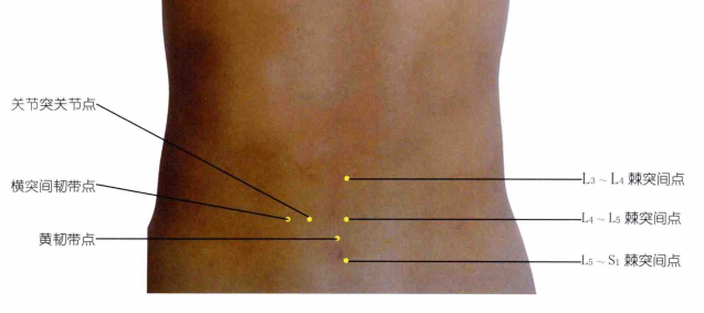
2. Treatment: small needle knife treatment can be given. The selected points were lumbar 4 / 5, lumbar 5 / sacral 1 interspinous point, transverse process point, articular process point and piriformis muscle point. After treatment, the patient's symptoms were slightly relieved, and the pain at night was reduced to VAS 6 points.
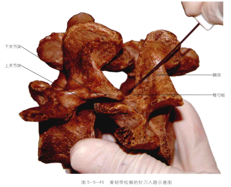
3. On the 3rd day after admission, improve the lumbar spine MR prompt: the lumbar 5 / sacral 1 intervertebral disc plane can see the right posterior lateral protrusion of the intervertebral disc, thickening of the ligamentum flavum, hypertrophy of the articular process, narrowing of the lateral recess, and nerve root compression. According to the clinical symptoms and MR results, it was considered as lumbar lateral recess stenosis. Adjust the treatment plan, and increase the lateral recess point (release the ligamentum flavum) and the external opening point of the intervertebral foramen at the needle knife treatment point.

Needle knife treatment point

Schematic diagram of ligamentum flavum release
Treatment Process
After three times of treatment, the leg pain of the patient's right lower limb was basically relieved, and the VAS score was reduced to 1-2 points. In the later stage, the core muscle group training and self stretching training were taught to improve the stability of the spine and prevent the recurrence of low back pain.

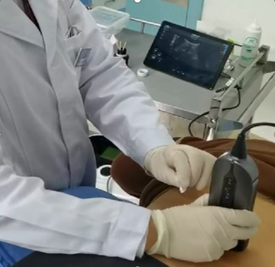
SonoEye for guiding needle knife treatment of lumbar disc herniation
Summary
1. This case belongs to the special lumbar disc herniation - lumbar lateral recess stenosis. According to MR film, the lumbar intervertebral disc protrusion is not particularly serious, but the protruding part is just at the lateral recess, resulting in obvious nerve root compression and severe clinical symptoms.
2.The lumbar lateral recess is located at the inner side of the pedicle, with the superior articular process and the ligamentum flavum at the rear, and the intervertebral disc and vertebral body at the front and outside. It opens inward to the vertebral canal and continues outward to the intervertebral canal. The spinal canal of the fifth lumbar vertebra is trilobal, and its lateral recess is the narrowest. Therefore, the deformation and protrusion of the intervertebral disc, the labial hyperplasia of the posterior edge of the vertebral body, the hyperplasia and hypertrophy of the lower joint, and the thickening of the ligamentum flavum can all lead to the stenosis of the lateral recess. The anteroposterior diameter of the lateral recess of this patient is only 2mm, which belongs to severe stenosis.
3. The stenosis of the lumbar lateral recess can lead to untreatable pain, which is severe. The pain cannot be alleviated by general pain relievers, and the symptoms cannot be alleviated by lying flat. It needs to be alleviated by bending and standing or bending the hips and knees on the side. At this time, the lumbar spine is in the flexion position, the ligamentum flavum is tense and thin, the upper articular process does not move forward, the space of the lateral recess is relatively large, and the nerve pressure will be reduced. This also suggests that the release of ligamentum flavum is the key point of our needle knife treatment. In addition, the patient's lower limb radiation pain was severe, but there was no obvious low back pain, indicating that the anterior branch of the nerve root was compressed and the posterior branch was not affected. This suggests that the external opening of the intervertebral foramen can be loosened, which may relieve the compression of the nerve root. However, the positions of the above two anatomical sites are deep, and the needle path of the needle knife is limited. The success rate of blind penetration to the target point is very low. In this case, SonoEye handheld ultrasound-guided needle knife is used in the guided procedure which can accurately penetrate the needle tip to the treatment site, quickly, accurately and efficiently, and avoid the damage caused by repeated blind puncture.

4. The degenerative narrowing of the lumbar 5 / sacral 1 intervertebral disc of the patient indicates that the intervertebral disc bears great vertical pressure. Besides the body weight of the upper body, the pressure also comes from the muscles, ligaments and joint capsules around the intervertebral disc, including supraspinous / interspinous ligaments, intertransverse muscles and ligaments, psoas major muscle, erector spinalis muscle and joint capsule of the articular process. These are all the treatment points of needle knife. These treatment points are relatively shallow, with bony reference points and no large neurovascular organs around them, so they can be punctured by ultrasound guidance or by hand.
Contact Us
Copyright © CHISON Medical Technologies Co., Ltd. All Rights Reserved | Privacy and terms of use
Products: Vascular Ultrasound Machine MSK Ultrasound Machine Portable Ultrasound Device Portable Ultrasound Machine for Sale Portable Ultrasound Machine For Pregnancy Handheld Ultrasound For Pregnancy Handheld Veterinary Ultrasound Veterinary Ultrasound Machine Portable Vascular Ultrasound Livestock Ultrasound Machine

CHISON respects your privacy. We use cookies to make our site more personal and enhance your experience. Read our Private Policy to learn more about cookies and how to manage them. You agree to our use of these technologies when you visit our site.

We appreciate your feedback
We sincerely invite you to participate in our survey for helping us to improve our digital market.
*1.How fast does the website load?
*2.Does the products displayed on the website interest you?
*3.How easy is it for you to find the information you need?
4.What information or service do you suggest we can offer?


THANKS
Thank you for sharing your thoughts with us. We’re highly appreciated your every feedback.

Sorry!

THANKS
Thank you for sharing your thoughts with us. We’re highly appreciated your every feedback.