


Clinical Insights
Thyroid ultrasound examinations are often centered around one primary question:
“Is there a thyroid nodule?”
However, not all thyroid diseases present as focal lesions. In daily clinical practice, diffuse thyroid disease can be far more subtle — and far easier to overlook during routine scanning.
This case highlights how careful evaluation of thyroid parenchyma, vascularity, and overall glandular appearance can provide important diagnostic clues, even in the absence of discrete nodules.
A patient presented with anterior neck discomfort and abnormal thyroid-related symptoms. Laboratory evaluation suggested thyroid dysfunction, and ultrasound examination was requested for further assessment.
The examination was performed using the SonoMax ultrasound system with a high-frequency linear transducer.

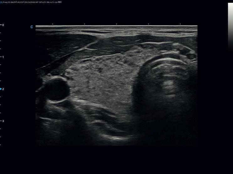
Grey-scale ultrasound demonstrated diffusely heterogeneous thyroid parenchyma with multiple patchy and cord-like hypoechoic areas throughout the gland. The glandular echotexture appeared coarse, suggesting diffuse parenchymal involvement.
No well-defined focal nodule with a clear margin or complete capsule was identified. Instead, the hypoechoic regions blended gradually into the surrounding tissue rather than forming discrete focal lesions.
These findings were suggestive of diffuse thyroid parenchymal disease.
Color Doppler imaging demonstrated increased vascularity within the thyroid parenchyma, with punctate and patchy flow signals diffusely distributed throughout the gland.
The vascularity was not confined to the periphery of a focal lesion but involved the thyroid diffusely, suggesting globally increased glandular perfusion.
Together, these findings were highly suggestive of diffuse inflammatory thyroid disease.
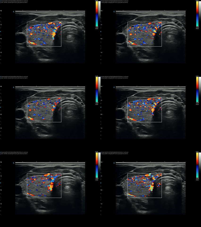
Color Doppler imaging
Longitudinal Imaging and Volume Flow Assessment
On longitudinal imaging, the thyroid parenchyma appeared diffusely coarse and heterogeneous, with intermixed hypoechoic and relatively hyperechoic regions creating a disorganized glandular texture.
Volume Flow imaging demonstrated abundant intrathyroidal vascular signals with patchy, strip-like, and branching patterns. These vascular findings suggested active perfusion within the thyroid vascular bed.
Importantly, these findings were consistently observed across multiple longitudinal scanning planes rather than representing isolated changes on a single image, further supporting diffuse glandular involvement.

High‑Sensitivity Flow imaging
Magnified Vascular Assessment
Magnified imaging further demonstrated vascular signals distributed within the thyroid parenchyma and adjacent peripheral regions.
Some vessels appeared as continuous strip-like and branching flow patterns, providing additional evidence of diffuse hypervascularity associated with inflammatory thyroid disease.
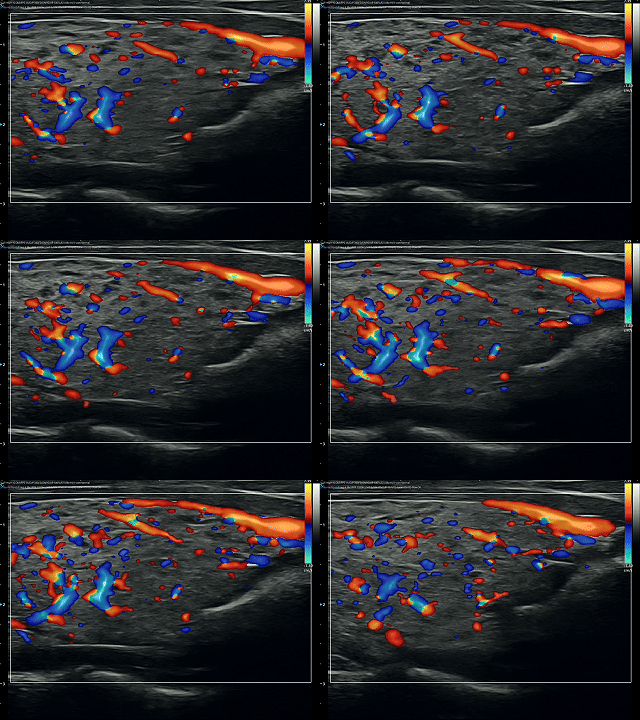
Magnified Vascular Assessment
In routine thyroid imaging, the absence of nodules can sometimes lead to underestimation of diffuse glandular abnormalities.
In this case, several ultrasound findings were particularly important:
l Diffuse heterogeneous echotexture
l Ill-defined “map-like” hypoechoic regions
l Absence of discrete nodules
l Increased intrathyroidal vascularity
These findings shifted the diagnostic focus away from focal thyroid lesions and toward diffuse thyroid pathology.
Based on the imaging appearance and clinical presentation, the findings were considered highly suggestive of diffuse inflammatory thyroid disease
In routine thyroid ultrasound examinations, attention is often directed toward identifying focal nodules. As a result, diffuse parenchymal abnormalities may receive less attention, particularly in patients without obvious mass-like lesions.
This case highlights the importance of evaluating:
l Overall glandular echotexture
l Distribution of hypoechoic changes
l Vascular patterns on Doppler imaging
l Correlation with clinical symptoms and laboratory findings
A comprehensive ultrasound assessment can provide valuable diagnostic direction and help guide further endocrine evaluation.
l Not all thyroid disease presents as nodules
l Diffuse “map-like” hypoechoic changes are important diagnostic clues
l Increased Doppler flow may indicate active inflammatory involvement
l Careful assessment of diffuse parenchymal abnormalities can improve diagnostic confidence
l Ultrasound plays a key role in the early evaluation of diffuse thyroid disease
This case serves as a reminder that thyroid ultrasound should not be limited to nodule detection alone.
In diffuse thyroid disease, subtle changes in echotexture and vascularity may provide critical insights into underlying inflammatory processes and support earlier clinical diagnosis.
Cases like this reinforce the value of systematic thyroid evaluation and the importance of looking beyond focal lesions during routine ultrasound examinations.
1.Vitti P, Rago T, Mazzeo S, et al. Thyroid blood flow evaluation by color-flow Doppler sonography distinguishes Graves’ disease from Hashimoto’s thyroiditis. J Endocrinol Invest. 1995;18(11):857–861.
2.Pedersen OM, Aardal NP, Larssen TB, et al. The value of ultrasonography in predicting autoimmune thyroid disease. Thyroid. 2000;10(3):251–259.
3.Rago T, Chiovato L, Grasso L, et al. Thyroid ultrasonography as a tool for detecting thyroid autoimmune diseases and predicting thyroid dysfunction in apparently healthy subjects. J Endocrinol Invest. 2001;24(10):763–769.
4.Dietrich CF, Müller T, Bojunga J, et al. Ultrasound of the Thyroid. European Federation of Societies for Ultrasound in Medicine and Biology Recommendations. Ultrasound Int Open. 2017;3(1):E1–E26.
This article is intended for educational and professional communication purposes only. Certain reference materials and images were adapted from publicly available sources. Clinical decisions should be made based on comprehensive medical evaluation by qualified healthcare professionals.

Contact Us
Copyright © CHISON Medical Technologies Co., Ltd. All Rights Reserved | Privacy and terms of use
Products: Vascular Ultrasound Machine MSK Ultrasound Machine Portable Ultrasound Device Portable Ultrasound Machine for Sale Portable Ultrasound Machines For Pregnancy Handheld Ultrasound For Pregnancy Handheld Veterinary Ultrasound Veterinary Ultrasound Machine Portable Vascular Ultrasound Livestock Ultrasound Machine

CHISON respects your privacy. We use cookies to make our site more personal and enhance your experience. Read our Private Policy to learn more about cookies and how to manage them. You agree to our use of these technologies when you visit our site.
We appreciate your feedback
We sincerely invite you to participate in our survey for helping us to improve our digital market.
*1.How fast does the website load?
*2.Does the products displayed on the website interest you?
*3.How easy is it for you to find the information you need?
4.What information or service do you suggest we can offer?

THANKS
Thank you for sharing your thoughts with us. We’re highly appreciated your every feedback.
Sorry!
THANKS
Thank you for sharing your thoughts with us. We’re highly appreciated your every feedback.
