



Clinical Insights
CHISON | SonoMax Ultrasound Case Share
For professional education and communication only. Not a substitute for clinical diagnosis.
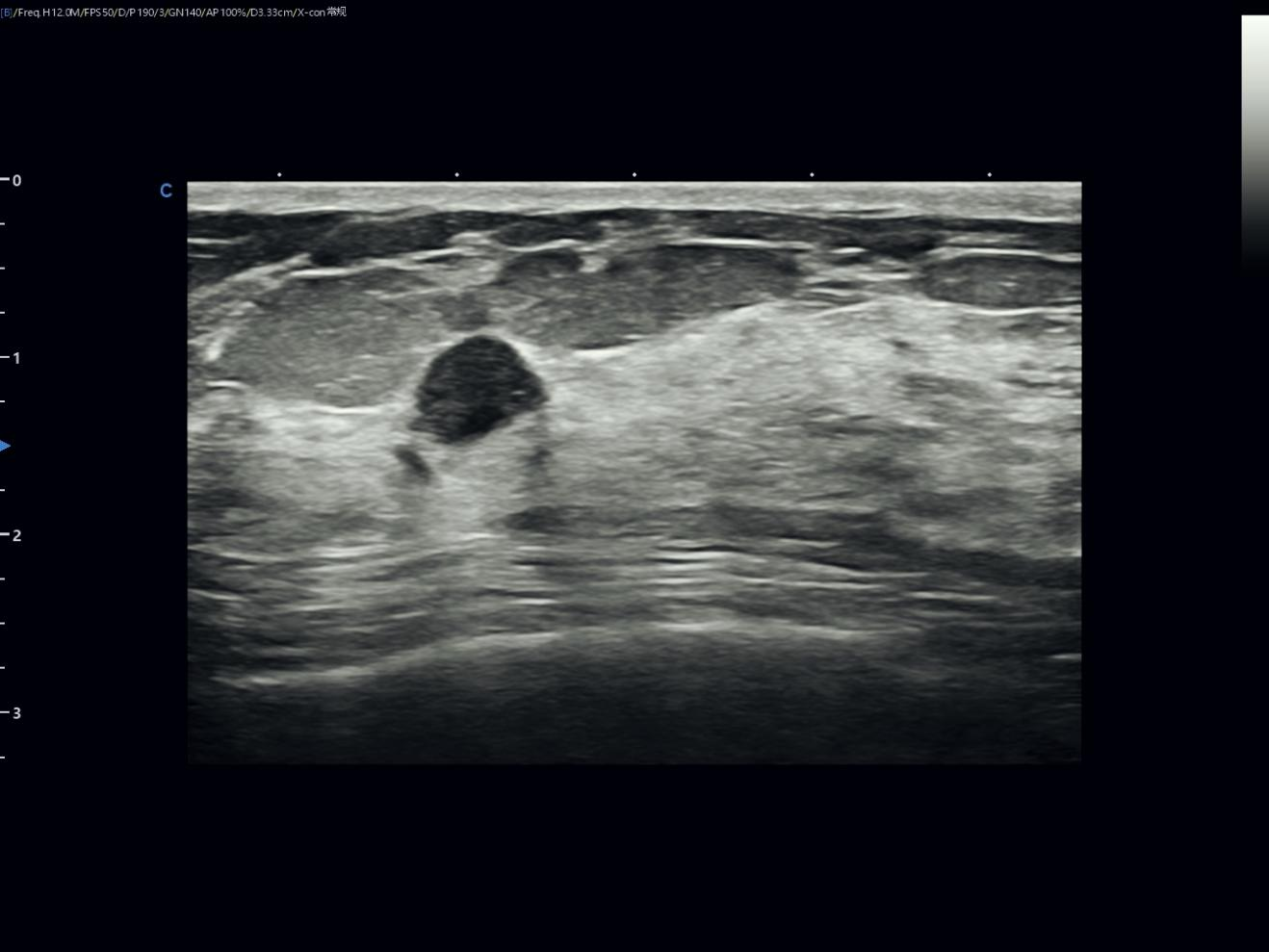
In this case, high-frequency ultrasound of the breast demonstrated a small solid hypoechoic nodule within the breast parenchyma. The lesion was relatively well circumscribed, oval to round in shape, and showed no obvious suspicious vascularity on the provided Q-flow /color Doppler images.
Taken together, the imaging appearance was most consistent with breast fibroadenoma.
Ultrasound system: SonoMax
Probe: L5-14 high-frequency linear probe
Examination region: Breast
Imaging modes: Grey-scale ultrasound, Q-flow / color Doppler assessment
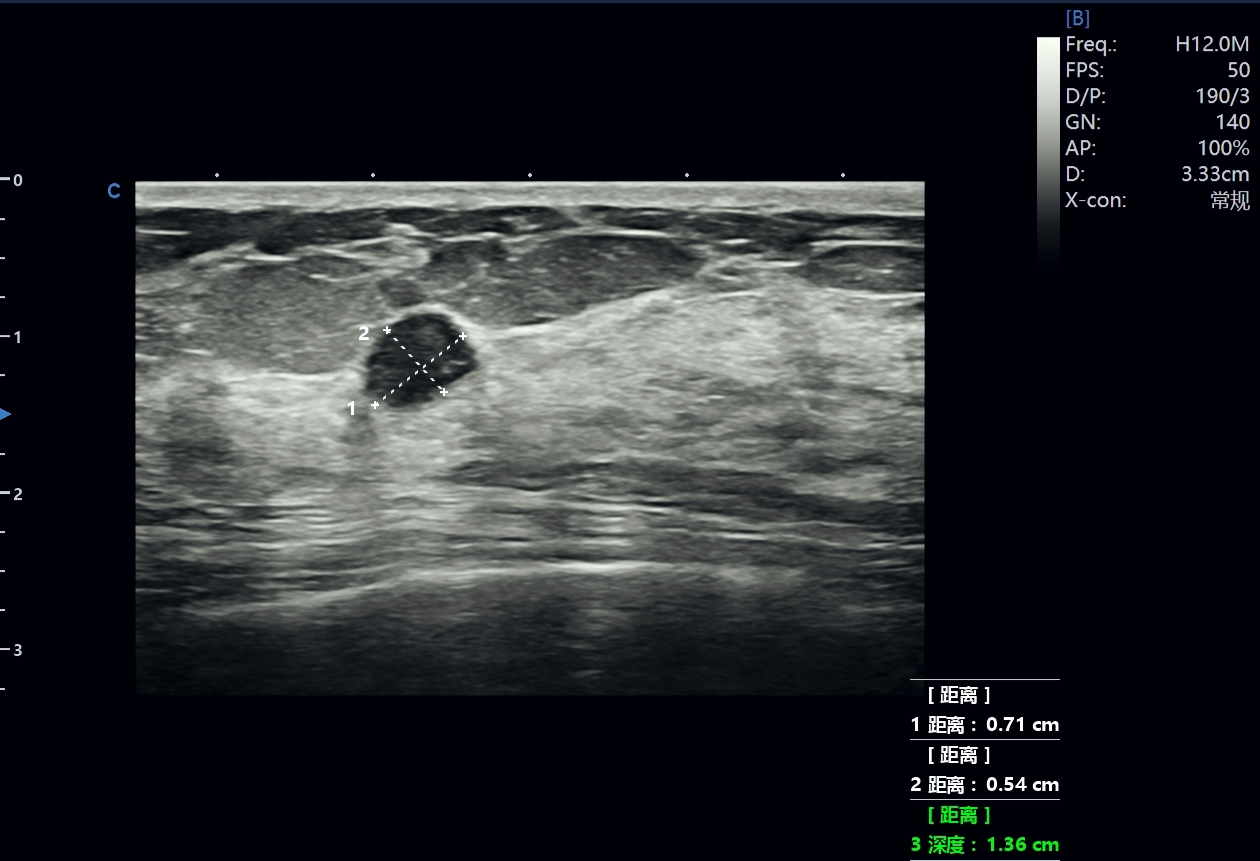
Across multiple grey-scale and Doppler views, ultrasound showed a small solid hypoechoic nodule within the breast parenchyma. The target lesion measured approximately 0.71 cm × 0.54 cm, with a recorded depth of approximately 1.36 cm from the skin surface.
The lesion appeared oval to round, relatively well defined, and internally homogeneous. No definite posterior acoustic shadowing, architectural distortion, suspicious calcification, or marked internal vascularity was identified in the provided images.
The overall imaging pattern supported the impression of a benign-appearing solid breast nodule, consistent with fibroadenoma. [1,2,3]
Figure 1. Grey-Scale Ultrasound Showing a Solid Hypoechoic Breast Nodule
High-frequency grey-scale ultrasound showed clear visualization of the superficial breast layers and breast parenchyma. Within the breast tissue, a small solid hypoechoic nodule was identified, measuring approximately0.71 cm × 0.54cm, with a recorded depth of approximately1.36cm from the skin surface. The lesion appeared oval to round, relatively well circumscribed, and internally homogeneous, without a definite taller-than-wide configuration. No obvious cystic component, coarse calcification, posterior acoustic shadowing, tissue retraction, or surrounding architectural distortion was seen in the provided images. Overall, these grey-scale and measurement findings supported a benign-appearing solid breast nodule, compatible with the common sonographic appearance of fibroadenoma when correlated with the patient’s clinical background and follow-up status. [1,2,3]
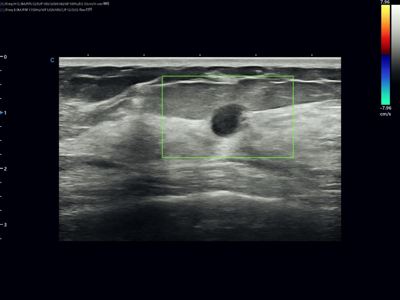
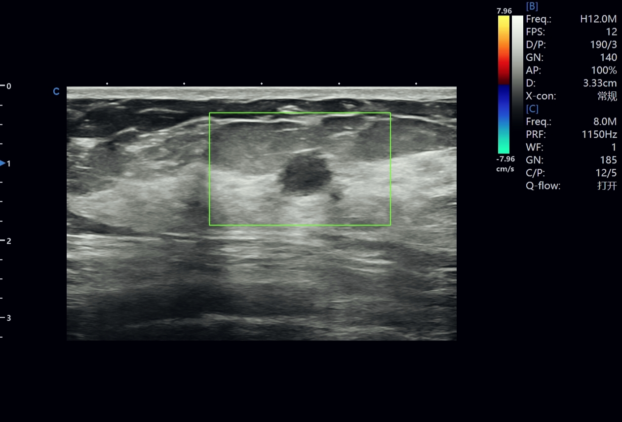
Q-flow / color Doppler imaging demonstrated the target nodule within the assessment box. The lesion remained a solid hypoechoic nodule with an oval-to-round shape and a relatively clear boundary. In the provided static images, no definite rich internal vascularity, abnormal penetrating vessel, chaotic vascular pattern, or prominent peripheral vascular ring was identified. Together with the lesion’s regular morphology and relatively homogeneous internal echo pattern on grey-scale ultrasound, the absence of marked suspicious vascularity supported a benign-leaning imaging impression. Doppler findings should be interpreted as complementary to grey-scale morphology rather than as a standalone diagnostic criterion. [5]
This finding was consistent with a benign-appearing solid breast nodule, while clinical correlation and BI-RADS-based assessment remain necessary.
Grey-scale survey images demonstrated the breast parenchyma surrounding the target nodule, with recognizable fibroglandular tissue, fatty lobules, echogenic fibrous septa, and deeper fascial structures. Across the provided views, no definite additional suspicious solid mass, ductal dilatation, focal fluid collection, abnormal posterior acoustic shadowing, architectural distortion, skin involvement, or obvious suspicious change directly associated with the target lesion was identified. These images provided background information on the surrounding breast tissue and supported lesion localization, contextual assessment, and follow-up comparison together with the target nodule images.
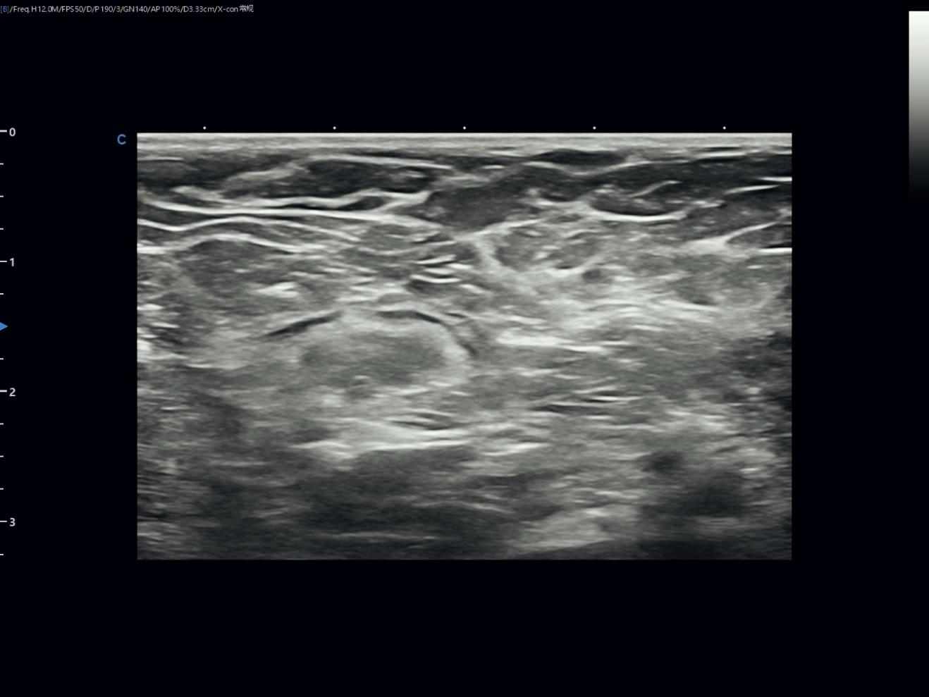
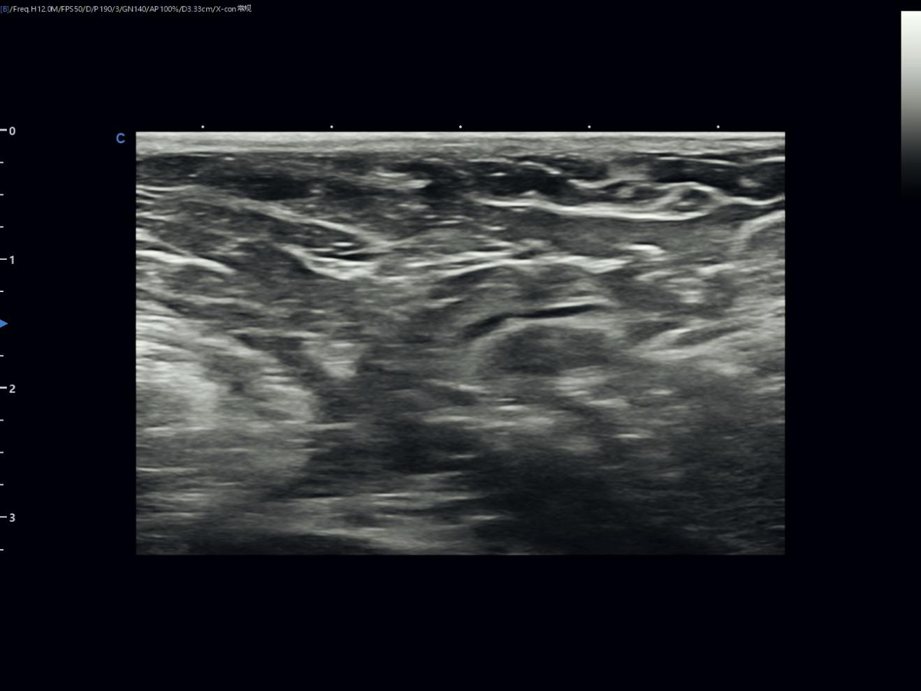
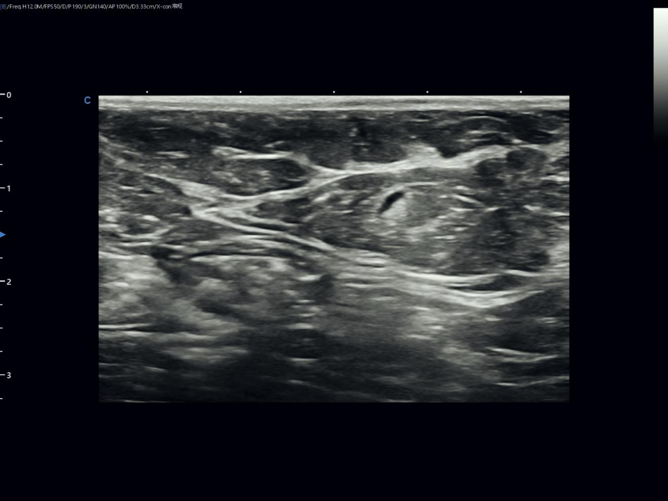
Breast fibroadenoma is one of the most common benign solid breast lesions. It is a fibroepithelial lesion composed of epithelial and stromal components and is frequently encountered in young women, although it may be detected across a broad age range.
On ultrasound, fibroadenomas often appear as oval, circumscribed, parallel-oriented, homogeneous hypoechoic solid masses. Some may show gentle lobulation, a thin echogenic capsule, or mild posterior acoustic enhancement. [1,2]
In this case, the most important imaging features were:
A small solid hypoechoic breast nodule
A measured size of approximately 0.71 cm × 0.54 cm
A relatively clear margin
Oval to round morphology
Relatively homogeneous internal echo pattern
No definite taller-than-wide configuration
No obvious posterior acoustic shadowing
No obvious suspicious calcification
No clear rich internal vascularity on Q-flow / Doppler images
No associated architectural distortion in the provided views
The overall imaging impression is therefore most consistent with:
A benign-appearing solid breast nodule, compatible with breast fibroadenoma.
This interpretation is not based solely on the fact that the lesion is small. It is based on a structured evaluation of its shape, margin, orientation, echo pattern, posterior features, vascularity, and relationship with surrounding breast tissue.
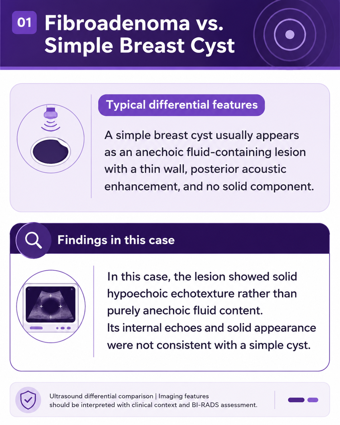
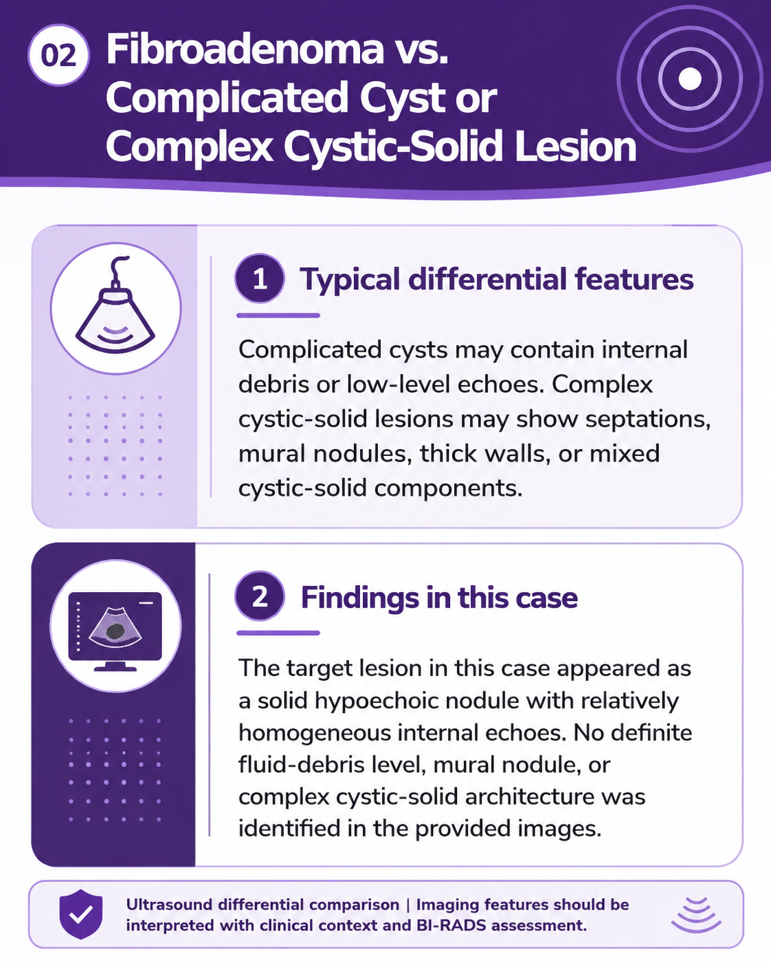
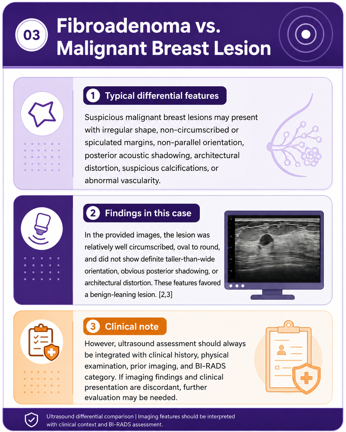
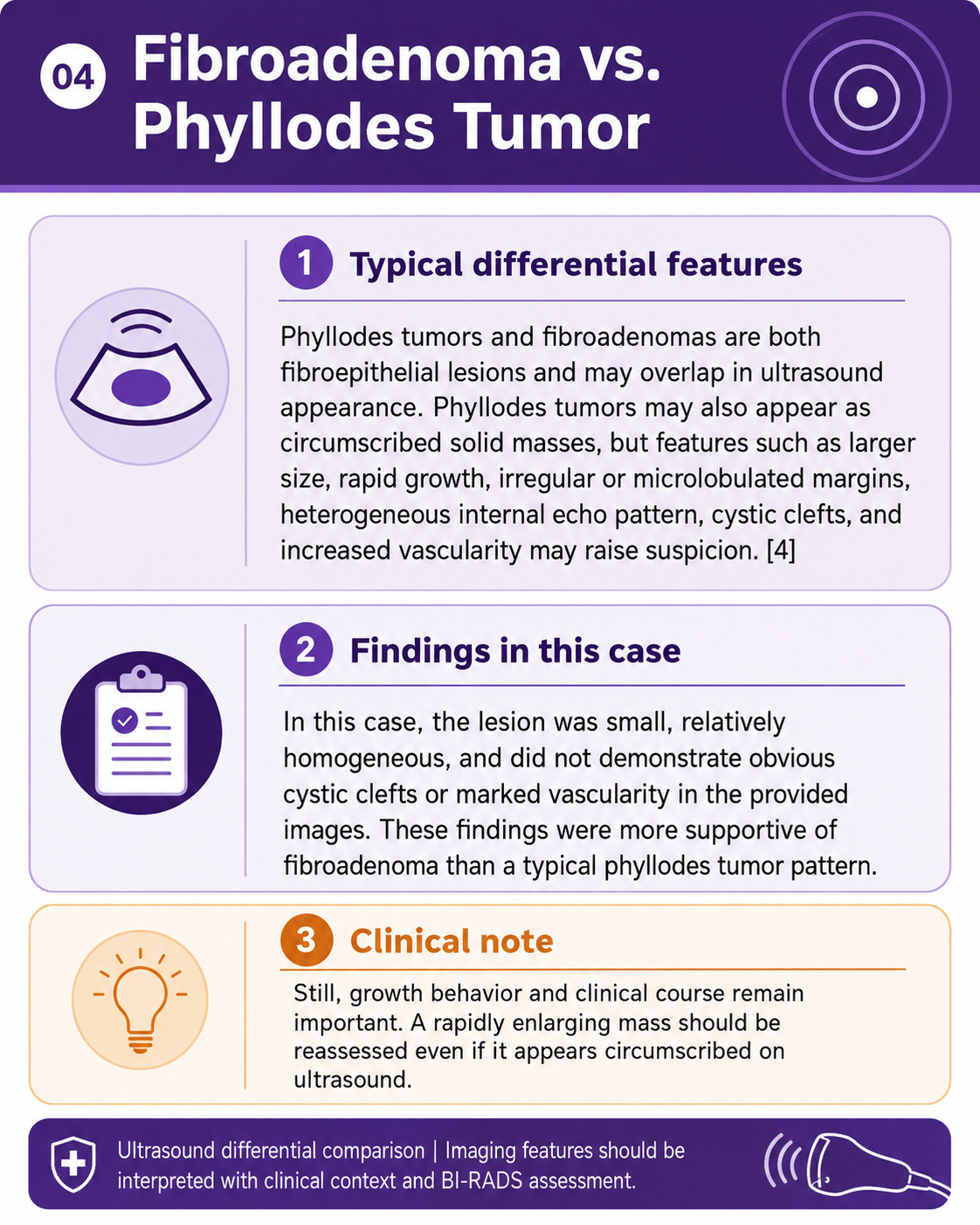
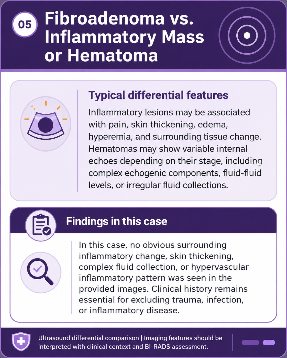
A small breast nodule should be evaluated by composition, shape, orientation, margin, echo pattern, posterior acoustic features, calcifications, vascularity, and associated findings.
Fibroadenoma commonly appears as an oval, circumscribed, parallel-oriented, homogeneous hypoechoic solid mass. [1,2]
Color Doppler or Q-flow findings are useful but complementary. The absence of marked vascularity supports a benign-leaning impression, but grey-scale morphology remains central to assessment. [5]
Fibroadenoma and phyllodes tumor may overlap in imaging appearance. Rapid growth, larger size, heterogeneous echo pattern, cystic clefts, microlobulated margins, or increased vascularity should prompt more careful evaluation. [4]
The lesion in this case measured approximately 0.71 cm × 0.54 cm, appeared relatively well circumscribed and homogeneous, and showed no obvious suspicious vascularity in the provided images.
The overall imaging appearance supports the impression of breast fibroadenoma.
A breast fibroadenoma may appear as a small, well-defined nodule. But ultrasound interpretation should never stop at “a small mass was found.”
By showing the lesion’s solid composition, shape, margin, internal echo pattern, vascularity, and relationship with surrounding breast tissue, ultrasound helps clinicians move from detection toward interpretation.
With high-frequency, clear, and reliable imaging, SonoMax supports clinicians in identifying subtle breast findings, documenting meaningful lesion characteristics, and making each examination more valuable for patient care.
Great sound is often quiet. True vision begins in the subtle.
CHISON remains committed to advancing ultrasound innovation, creating clinical value, and helping make healthcare more accessible and precise.
1. Masciadri N, Ferranti C. Benign breast lesions: Ultrasound. Journal of Ultrasound. 2011;14(2):55-65. doi:10.1016/j.jus.2011.03.002.
2. Stavros AT, Thickman D, Rapp CL, Dennis MA, Parker SH, Sisney GA. Solid Breast Nodules: Use of Sonography to Distinguish between Benign and Malignant Lesions. Radiology. 1995;196(1):123-134.
3. Lee J, Lee JH, Baik S, et al. Practical and Illustrated Summary of Updated BI-RADS for Ultrasonography. Ultrasonography. 2017;36(1):71-81. doi:10.14366/usg.16034.
4. Duman L, Gezer NS, Balcı P, Altay C, Başara I, Durak MG, Sevinç AI. Differentiation between Phyllodes Tumors and Fibroadenomas Based on Mammographic, Sonographic and MRI Features. Breast Care. 2016;11(2):123-127. doi:10.1159/000444377.
5. Busilacchi P, Draghi F, Preda L, et al. Has Color Doppler a Role in the Evaluation of Mammary Lesions? Journal of Ultrasound. 2012;15(2):93-98. doi:10.1016/j.jus.2012.02.002.
Note: This case is intended for professional education and communication only. It is not a substitute for clinical diagnosis or treatment. If symptoms occur, patients should seek care from qualified medical professionals.

Contact Us
Copyright © CHISON Medical Technologies Co., Ltd. All Rights Reserved | Privacy and terms of use
Products: Vascular Ultrasound Machine MSK Ultrasound Machine Portable Ultrasound Device Portable Ultrasound Machine for Sale Portable Ultrasound Machines For Pregnancy Handheld Ultrasound For Pregnancy Handheld Veterinary Ultrasound Veterinary Ultrasound Machine Portable Vascular Ultrasound Livestock Ultrasound Machine

CHISON respects your privacy. We use cookies to make our site more personal and enhance your experience. Read our Private Policy to learn more about cookies and how to manage them. You agree to our use of these technologies when you visit our site.
We appreciate your feedback
We sincerely invite you to participate in our survey for helping us to improve our digital market.
*1.How fast does the website load?
*2.Does the products displayed on the website interest you?
*3.How easy is it for you to find the information you need?
4.What information or service do you suggest we can offer?

THANKS
Thank you for sharing your thoughts with us. We’re highly appreciated your every feedback.
Sorry!
THANKS
Thank you for sharing your thoughts with us. We’re highly appreciated your every feedback.
