



Clinical Insights
Fetal Lateral Ventricle Dilatation on Prenatal Ultrasound
SonoMax | Prenatal Ultrasound | Fetal Neurosonography
Ultrasound in pregnancy plays an essential role in assessing fetal growth and anatomy. This prenatal ultrasound case focuses on localized cystic dilatation of one fetal lateral ventricle and explains why the finding must be distinguished from a standardized ventriculomegaly assessment.
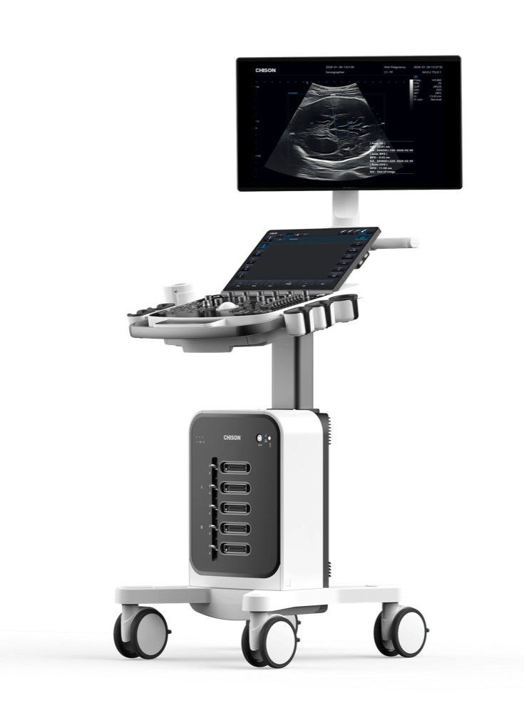
Ultrasound system: SonoMax
Ultrasound findings: A transverse view of the fetal brain shows a continuous hyperechoic calvarial ring with visible brain parenchyma. A localized, marked cystic dilatation is seen in one lateral ventricle. The dilated area appears as an anechoic, fluid-filled space with a relatively regular shape and well-defined margins. Dynamic scanning helps confirm that the fluid-filled space persists and allows its spatial relationship to the lateral ventricle and surrounding brain tissue to be assessed from different insonation angles.
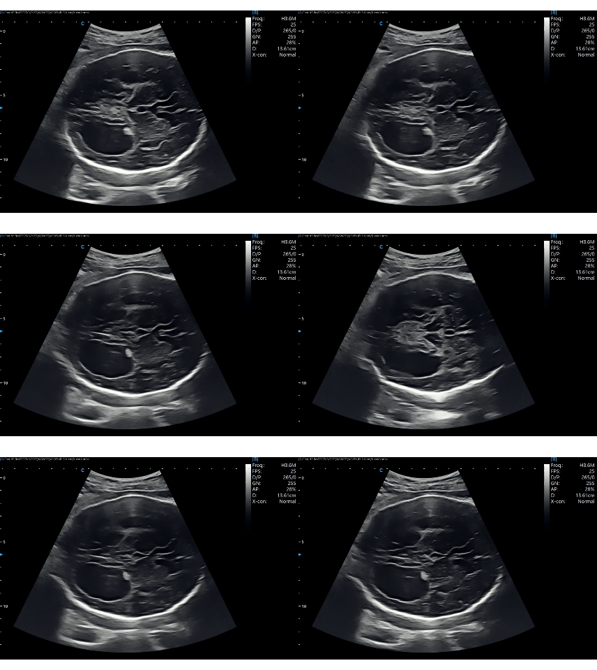
Figure 1. Localized anechoic area in one fetal lateral ventricle.
Ultrasound findings: Two-dimensional grayscale imaging further demonstrates enlargement in the region of one lateral ventricle, with predominantly anechoic fluid content. The cystically dilated area has a clear outline; the adjacent brain parenchyma remains distinguishable, and the posterior calvarial echo is continuous. In this plane, the fluid-filled area lies within the anatomic region of the lateral ventricle, and its morphology and location are consistent with localized cystic dilatation of the lateral ventricle.
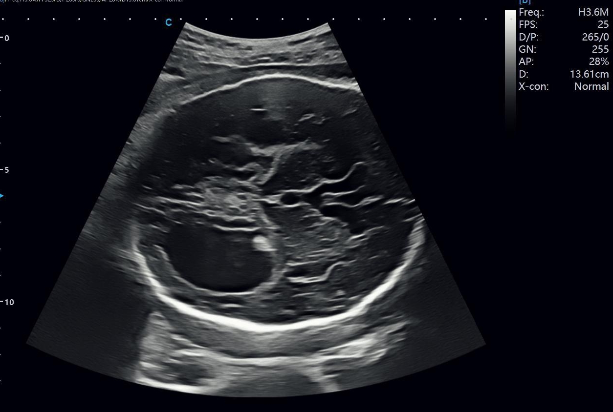
Figure 2. Two-dimensional grayscale view of the localized cystically dilated ventricular region.
Ultrasound findings: On a clearly visualized transverse view of the fetal brain, a localized diameter of the cystically dilated lateral ventricular region was measured at approximately 2.74 cm.
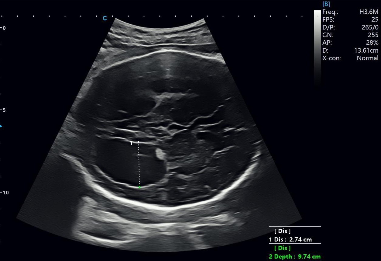
Figure 3. Localized diameter measurement of approximately 2.74 cm; this is not the standardized atrial width.
The system automatically identified the fetal calvarial contour, cerebral midline, and relevant intracranial landmarks, then completed measurements of head circumference (HC), biparietal diameter (BPD), and occipitofrontal diameter (OFD). The automated results were: HC approximately 33.61 cm, corresponding to 38 weeks 3 days; BPD approximately 9.53 cm, corresponding to 38 weeks 6 days; and OFD approximately 11.50 cm. Automated fetal head biometry can provide quantitative support for fetal growth assessment. However, evaluation of lateral ventricular cystic dilatation must still be based on standardized planes, dynamic multiplanar scanning, and the physician's integrated interpretation of the anatomy.
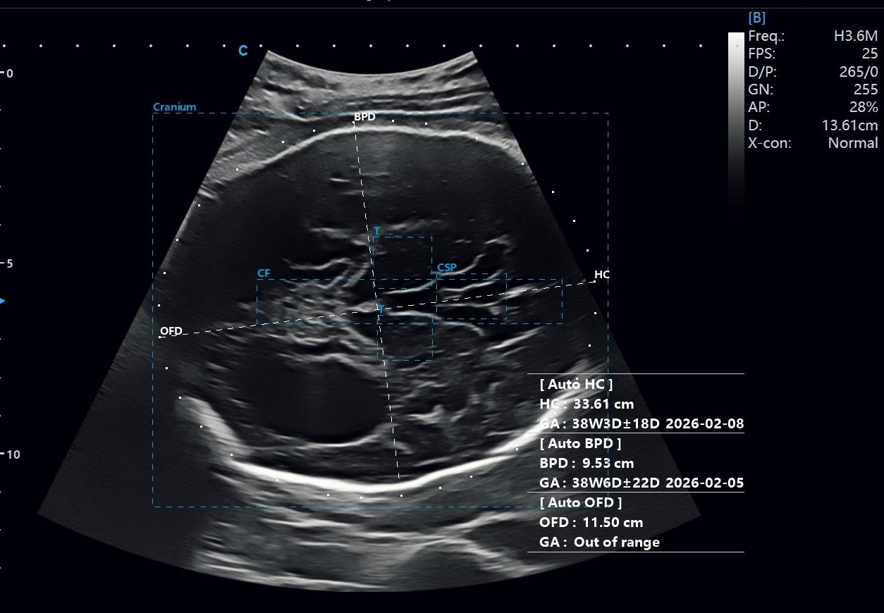
Figure 4. Automated fetal head biometry, including HC, BPD, and OFD measurements.
The fetal lateral ventricles are part of the cerebrospinal fluid circulation system. When prenatal ultrasound shows fluid-filled dilatation in this region, the examiner should confirm that the anechoic area is intraventricular and determine whether it is unilateral or bilateral, focal or diffuse, and stable or progressive.
ISUOG recommends evaluating the fetal central nervous system in the transventricular, transthalamic, and transcerebellar planes. If a structural abnormality is suspected, targeted fetal neurosonography should assess the ventricles, midline structures, corpus callosum, posterior fossa, and brain parenchyma in axial, coronal, and sagittal planes.
Interpretation requires more than identifying an anechoic area. Its relationship to the lateral ventricle, choroid plexus, ventricular wall, surrounding brain tissue, third and fourth ventricles, and posterior fossa should also be evaluated.
A localized measurement of the cystically dilated area is not equivalent to the standard atrial width of the lateral ventricle. The atrium should be measured at its widest point in a standard transventricular plane, using an inner-edge-to-inner-edge technique. Only a standardized atrial measurement should be used to assess lateral ventriculomegaly severity or prognosis.
Lateral ventricular abnormalities may be associated with impaired cerebrospinal fluid circulation, central nervous system malformations, intrauterine infection, hemorrhage, or genetic abnormalities. Their clinical significance depends on the degree and progression of dilatation and the presence of associated abnormalities.
This case shows a well-defined, anechoic, fluid-filled area within the anatomic region of one lateral ventricle. Multiplanar imaging supports localized lateral ventricular cystic dilatation. Differential diagnosis depends mainly on the lesion’s relationship to the ventricular system, brain parenchyma, and extra-axial spaces.
Porencephalic or encephalomalacic cavities are located within the brain parenchyma and may be associated with loss of adjacent brain tissue. They may also communicate with the ventricle or subarachnoid space. Lateral ventricular cystic dilatation is primarily intraventricular and should be assessed using the choroid plexus position and multiplanar continuity.
An arachnoid cyst is an extra-axial fluid-filled lesion that may compress adjacent brain tissue or ventricles. In contrast, lateral ventricular cystic dilatation remains within the anatomic boundaries of the ventricle.
A choroid plexus cyst usually appears as a small anechoic area surrounded by echogenic choroid plexus tissue. In this case, the larger fluid-filled area represents dilatation of the lateral ventricular cavity rather than a cyst confined to the choroid plexus.
Hydrocephalus may involve progressive ventricular enlargement caused by abnormal cerebrospinal fluid production, circulation, or absorption. The available images show only localized cystic dilatation of one lateral ventricle. Without serial examinations, bilateral ventricular measurements, and a complete central nervous system assessment, hydrocephalus cannot be confirmed.
After detecting a fetal lateral ventricular abnormality, a targeted MFM scan or fetal neurosonography should assess both lateral ventricles, the third and fourth ventricles, cavum septi pellucidi, corpus callosum, thalami, cortical development, posterior fossa, and spine. Serial ultrasound, fetal MRI, and infection or genetic testing may also be considered according to the complete findings and clinical history.
Two-dimensional ultrasound shows localized cystic dilatation of one fetal lateral ventricle. The area is anechoic and fluid-filled, with well-defined margins and a localized diameter of approximately 2.74 cm. These findings support the clinical interpretation of localized lateral ventricular cystic dilatation.
However, this localized measurement should not be interpreted as the standard atrial width. Dynamic scanning, standardized planes, and multiplanar assessment are required to determine the abnormality’s location, morphology, and relationship to surrounding brain tissue.
The available images cannot establish the cause, progression, or presence of associated central nervous system abnormalities. Complete fetal neurosonography and relevant clinical information are therefore required. Automated fetal head biometry can support routine HC and BPD measurements, but accurate diagnosis still depends on standardized scanning and professional clinical judgment.
CHISON remains committed to advancing healthcare through technological innovation and supporting better health for all.

Malinger G, Paladini D, Haratz KK, et al. ISUOG Practice Guidelines (updated): sonographic examination of the fetal central nervous system. Part 1: performance of screening examination and indications for targeted neurosonography. Ultrasound Obstet Gynecol. 2020;56(3):476-484. doi:10.1002/uog.22145.
Paladini D, Malinger G, Birnbaum R, et al. ISUOG Practice Guidelines (updated): sonographic examination of the fetal central nervous system. Part 2: performance of targeted neurosonography. Ultrasound Obstet Gynecol. 2021;57(4):661-671. doi:10.1002/uog.23616.
Fox NS, Monteagudo A, Kuller JA, Craigo S, Norton ME. Mild fetal ventriculomegaly: diagnosis, evaluation, and management. Am J Obstet Gynecol. 2018;219(1):B2-B9. doi:10.1016/j.ajog.2018.04.039.
Giorgione V, Haratz KK, Constantini S, Birnbaum R, Malinger G. Fetal cerebral ventriculomegaly: What do we tell the prospective parents? Prenat Diagn. 2022;42(13):1674-1681. doi:10.1002/pd.6266.
Alluhaybi AA, Altuhaini K, Alotaibi F, et al. Fetal ventriculomegaly: a review of literature. Cureus. 2022;14(2):e22152. doi:10.7759/cureus.22152.
Note: Some materials and images were compiled from publicly available sources. Please contact us regarding any copyright concerns. This content is for educational purposes only and does not replace professional medical consultation.

Contact Us
Copyright © CHISON Medical Technologies Co., Ltd. All Rights Reserved | Privacy and terms of use
Products: Vascular Ultrasound Machine MSK Ultrasound Machine Portable Ultrasound Device Portable Ultrasound Machine for Sale Portable Ultrasound Machines For Pregnancy Handheld Ultrasound For Pregnancy Handheld Veterinary Ultrasound Veterinary Ultrasound Machine Portable Vascular Ultrasound Livestock Ultrasound Machine

CHISON respects your privacy. We use cookies to make our site more personal and enhance your experience. Read our Private Policy to learn more about cookies and how to manage them. You agree to our use of these technologies when you visit our site.
We appreciate your feedback
We sincerely invite you to participate in our survey for helping us to improve our digital market.
*1.How fast does the website load?
*2.Does the products displayed on the website interest you?
*3.How easy is it for you to find the information you need?
4.What information or service do you suggest we can offer?

THANKS
Thank you for sharing your thoughts with us. We’re highly appreciated your every feedback.
Sorry!
THANKS
Thank you for sharing your thoughts with us. We’re highly appreciated your every feedback.
