Welcome to our comprehensive guide on shear wave elastography in ultrasound, a revolutionary technique that has transformed the field of diagnostic imaging. In this article, we will delve into the intricacies of shear wave elastography, exploring its principles, applications, and advantages over traditional imaging modalities. As experts in the field, we aim to provide you with valuable insights that will help you understand the significance of this cutting-edge technology.
Shear wave elastography is a non-invasive imaging technique that measures tissue stiffness by generating and analyzing shear waves within the body. By utilizing ultrasound technology, shear wave elastography offers a unique perspective into the mechanical properties of tissues, enabling healthcare professionals to diagnose a wide range of conditions accurately.

Shear wave elastography relies on the propagation of shear waves through tissues. These waves are generated by the application of acoustic radiation force, which causes displacements within the tissue. By measuring the speed at which these waves travel, shear wave elastography can determine the elasticity and stiffness of tissues. This information is then visualized using color maps, providing clinicians with valuable diagnostic information.
Shear wave elastography has gained significant traction in various medical specialties due to its versatility and accuracy. Let's explore some of its key applications:

Liver Disease Assessment
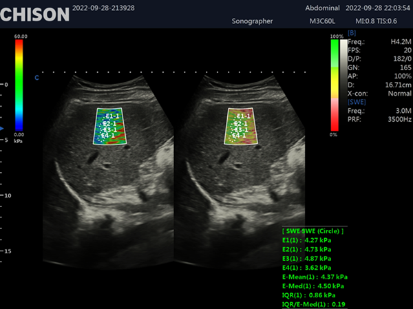
One of the most notable applications of shear wave elastography is in the assessment of liver disease. Traditional methods, such as liver biopsy, are invasive and carry certain risks. However, shear wave elastography provides a non-invasive alternative that can accurately quantify liver stiffness, aiding in the diagnosis and monitoring of conditions such as fibrosis and cirrhosis.
Breast Lesion Characterization
Shear wave elastography has shown immense promise in the field of breast imaging. By providing quantitative information about tissue stiffness, it helps differentiate between benign and malignant breast lesions. This aids in the early detection and diagnosis of breast cancer, allowing for timely intervention and improved patient outcomes.
Musculoskeletal Disorders
In the realm of musculoskeletal imaging, shear wave elastography has emerged as a valuable tool for assessing soft tissue injuries and disorders. It enables clinicians to precisely evaluate tendon and muscle elasticity, facilitating the diagnosis and monitoring of conditions like tendinopathy, muscle tears, and fibromyalgia.

Thyroid Nodule Assessment
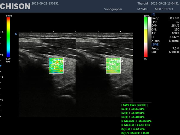
Shear wave elastography has also found application in the evaluation of thyroid nodules. Determining the stiffness of these nodules, it assists in distinguishing between benign and malignant lesions, reducing the need for unnecessary invasive procedures.
Compared to traditional imaging techniques, shear wave elastography offers several advantages that contribute to its growing popularity:
Non-Invasiveness
Shear wave elastography eliminates the need for invasive procedures such as biopsies in certain cases. This not only reduces patient discomfort but also minimizes associated risks and complications.
Real-Time Imaging
Unlike some imaging modalities, shear wave elastography provides real-time imaging capabilities. This allows healthcare professionals to assess tissue stiffness immediately during the examination, enabling prompt decision-making and enhancing patient care.
Quantitative Results
By providing quantitative measurements of tissue stiffness, shear wave elastography offers a level of objectivity that was previously unattainable. This facilitates accurate diagnosis, treatment planning, and monitoring of various conditions, leading to improved patient outcomes.
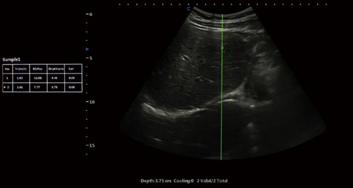
Shear wave elastography represents a significant advancement in the field of diagnostic imaging. With its ability to assess tissue stiffness non-invasively and provide valuable diagnostic information, this technique has revolutionized medical imaging across various specialties. From liver disease assessment to breast lesion characterization and musculoskeletal evaluations, shear wave elastography continues to enhance our understanding and management of numerous conditions. XBit 90 has both P-SWE point shear wave imaging and 2D-SWE surface shear wave imaging. Provide a variety of quantitative analysis parameters, such as velocity values, Young's modulus, and so on. Embracing this cutting-edge technology can empower healthcare professionals to deliver superior patient care and make more informed clinical decisions.
Contact Us
Copyright © CHISON Medical Technologies Co., Ltd. All Rights Reserved | Privacy and terms of use
Products: Vascular Ultrasound Machine MSK Ultrasound Machine Portable Ultrasound Device Portable Ultrasound Machine for Sale Portable Ultrasound Machine For Pregnancy Handheld Ultrasound For Pregnancy Handheld Veterinary Ultrasound Veterinary Ultrasound Machine Portable Vascular Ultrasound Livestock Ultrasound Machine

CHISON respects your privacy. We use cookies to make our site more personal and enhance your experience. Read our Private Policy to learn more about cookies and how to manage them. You agree to our use of these technologies when you visit our site.

We appreciate your feedback
We sincerely invite you to participate in our survey for helping us to improve our digital market.
*1.How fast does the website load?
*2.Does the products displayed on the website interest you?
*3.How easy is it for you to find the information you need?
4.What information or service do you suggest we can offer?


THANKS
Thank you for sharing your thoughts with us. We’re highly appreciated your every feedback.

Sorry!

THANKS
Thank you for sharing your thoughts with us. We’re highly appreciated your every feedback.